

Repairing Failing Dental Implants in Washington, DC
A loose, infected, or uncomfortable dental implant is evaluated for why it is failing and whether it can be saved before anything is removed. Specialist diagnosis in Washington, DC.
Understanding Why an Implant Is Failing Before Deciding What to Do
A dental implant that has become loose, tender, or surrounded by receding or bleeding gum tissue is not automatically lost. Implants fail for different reasons, and the reason determines the correct response. Some can be treated and kept. Some should be removed and replaced. Some are structurally sound but carry a restoration that was never designed to be cleaned or loaded properly, and the fix is a new crown rather than new surgery.
At Elite Prosthetic Dentistry, the evaluation of a failing implant answers four questions in order: why the implant is failing, whether it can be saved, what biological, structural, restorative, and bite-related factors are contributing, and what the appropriate correction is. The answer is reached from a clinical examination and a CBCT scan, and it is explained to you before any treatment is proposed. Patients are welcome whether the implant was placed here, by their own dentist, or at another practice, and many are referred by the dentist who continues to care for them.
How Implants Fail
Dental implants are the most durable tooth replacement available, but the tissues around them can become inflamed and infected in the same way the tissues around natural teeth can. Peri-implant mucositis is inflammation confined to the gum tissue; left untreated, it can progress to peri-implantitis, in which the bone supporting the implant is lost. Mechanical problems, such as a loose or fractured component, an implant placed at an angle the crown cannot compensate for, or an overloaded bite, add to the biological ones.
Dr. Marlin, a specialty-trained prosthodontist who has placed more than 3,900 dental implants over more than 40 years, evaluates both the surgical and the restorative side of a failing implant, because the two are usually connected.
What Causes Failing Implants?
From one tooth to an entire arch or to stabilize dentures, implant dentistry offers a successful solution for many restorative problems. While dental implants are the most reliable and only permanent tooth replacement solution, any of the following factors can result in peri-implantitis and failing implants.
Poor oral hygiene and untreated gum disease can cause bacterial infection around implant sites, increasing the risk of peri-implantitis and implant failure. As such, gum disease must be resolved before implant surgery, and proper oral health must be maintained after your procedure.
Implant position and planning matter. An implant placed where the final crown cannot be shaped and cleaned correctly, placed without adequate initial stability, or placed without accounting for the bite can lose bone over time even when the surgery itself healed well. Bone loss can also occur from excessive mechanical load on the implant.
Over time, various factors, including tooth loss, gum disease, and certain health conditions, can cause bone to lose density and weaken. Integration with the underlying bone is essential for implants to provide stable, long-lasting support for a crown, bridge, or full mouth prosthesis. If the underlying bone is inadequate, a bone grafting procedure is required to ensure proper fusion with the jaw bone.
Implant longevity depends on the surgical and restorative phases being planned together. Precision implant placement from a CBCT-based plan positions the implant to support the tooth that will sit on it, which reduces the risk of the mechanical and hygiene problems described above. Every mouth and every set of implants is different, which is why the evaluation of a failing implant starts with where it is and what it was asked to do.
It’s imperative to place an abutment and crown with the correct emergence profile (contour of the dental restoration where it meets the gum tissue). An over-contoured crown can make it difficult to clean, thereby increasing plaque growth/retention and the risk of gum inflammation.
Although the general health consequences of smoking are widely known, tobacco use increases the risk of several oral health issues. In terms of dental implants, research has shown a greater incidence of marginal bone loss, peri-implantitis, and implant failure, most notably in the grafted maxillary sinus area (upper back arch) in smokers.
Immune system disorders, steroid therapy, chemotherapy or radiosurgery in the neck area, and excessive bruxism (tooth clenching/grinding) can lead to dental implant failure. Although these issues don’t exclude candidacy, superb diagnostic and surgical skills are required to help ensure a successful outcome.
From Peri-Implant Mucositis to Peri-Implantitis
Peri-implant mucositis is inflammation and infection of the gum tissue surrounding a dental implant. Early symptoms include swollen gums, tenderness, and sensitivity. As the condition progresses to peri-implantitis, possible consequences include gum recession, deepening pockets, exposure of implant threads, drainage around the implant, and eventually a loose implant.
Treated early, peri-implantitis can often be managed by detoxifying the implant surface and grafting bone around it, without removing the implant.
Diagnosis Starts With Three-Dimensional Imaging
Every implant case at Elite Prosthetic Dentistry, from a single implant through full-mouth reconstruction, is planned on a CBCT (cone beam CT) scan. The scan shows bone volume and density, nerve and sinus anatomy, the space available for the restoration, and where the implant must sit to support the planned final tooth. This comprehensive and phased analysis is necessary to determine if your implant site requires a bone grafting procedure before implant placement. Surgical guides generated from the scan are used where bone anatomy, esthetic demands, or proximity to nerves and sinuses call for them. After the fusion process, our in-house lab technician creates the ideal, natural emergence profile without interference from misplaced implants.
Dr. Gerald M. Marlin was an early adopter of CBCT technology and has used CT-generated surgical guides since 2002. Since then, the technology has advanced from being merely preliminary viewing-analytic software to modern, interactive software allowing virtual reality “placement” of implant images in ideal positions on the CBCT 3D radiographic image for presurgical planning.
A Documented Example of Saving an Implant

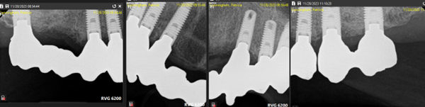
Treatment of a failing implant is based on the specific circumstances of that implant. As an example, one of our patients developed bone loss seven years after placement and restoration. To detoxify and re-graft the bone around a failing implant, the abutment and crown were removed, bone graft material was combined with a growth factor, and the implant was shielded under the protective gum tissue. Imaging after four months showed new bone formation that filled in around the side and top of the implant. After five months, the implant was exposed, and the abutment and crown were reseated, and the implant was saved without being removed.
Recognizing the Signs of Implant Trouble Early
Early recognition of implant problems dramatically improves the likelihood of successful treatment. Unfortunately, many patients do not seek evaluation until complications are advanced. Being aware of warning signs allows prompt intervention.
Bleeding or swollen gums around an implant are among the earliest signs of trouble. While some inflammation is normal immediately after placement, persistent inflammation beyond the first few weeks warrants evaluation. Increasing pocket depth (measured by probing around the implant) indicates advancing disease. Pus or drainage around the implant is a concerning sign of infection.
Some patients notice changes in implant mobility. A small amount of movement is normal immediately after placement and can occur when integration first occurs. But a tooth or implant that becomes progressively more mobile indicates compromised bone support and requires urgent evaluation.
Changes in bite or jaw position may indicate bone loss affecting implant support. Pain or discomfort around the implant, especially pain on biting, suggests a problem beneath the surface.
If you notice any of these signs, contact your dentist or our office promptly. Early evaluation and intervention often allow treatment that preserves the implant. Delayed diagnosis risks loss of the implant and additional bone loss that complicates replacement.
The Anatomy of Peri-Implantitis
To understand implant failure, one must understand peri-implantitis. Unlike tooth decay, which affects the tooth itself, peri-implantitis is an infection of the tissues and bone supporting the implant. It develops when bacteria colonize the space between the implant and the gum tissue, creating a pocket of infection similar to the periodontal pockets that develop around natural teeth with gum disease.
The infection extends progressively deeper, destroying gum tissue and bone around the implant. If untreated, the infection eventually undermines the implant’s support so completely that the implant becomes loose and fails.
What makes peri-implantitis particularly insidious is that it often develops without obvious symptoms until it is advanced. Unlike the acute pain of an abscessed natural tooth, peri-implantitis may cause no discomfort. A patient may not realize there is a problem until an implant becomes visibly loose or bleeding occurs.
This is why professional monitoring is so important. Even patients with excellent home care benefit from regular professional evaluation. Dr. Marlin’s examinations assess implant health, measure pocket depths, evaluate bone levels on radiographs, and identify early signs of trouble.
Risk Factors Beyond Surgical Technique
While surgical errors contribute to some implant failures, many failing implants resulted from proper surgery but developed problems afterward due to other factors. Understanding these risk factors helps patients understand what can be controlled and what requires vigilance.
Tobacco use is one of the most significant preventable risk factors. Smokers have significantly higher rates of implant failure and peri-implantitis. The mechanism is multiple: smoking impairs immune function, reduces blood supply to tissues, and promotes bacterial colonization. For smokers, implant success requires absolute commitment to excellent home care and more frequent professional monitoring.
Diabetes is another significant risk factor, particularly poorly controlled diabetes. Elevated blood glucose impairs immune function and bone healing. Diabetic patients benefit from more careful monitoring and sometimes more frequent professional cleanings.
Immune system disorders, including HIV infection and autoimmune conditions, affect implant success. Some medications, including certain steroids and chemotherapy agents, compromise healing and implant integration.
Bruxism (clenching and grinding) creates excessive mechanical forces on implants, potentially causing early bone loss. Patients with bruxism benefit from wearing a night guard to protect implants and natural teeth.
Oral hygiene is perhaps the most controllable risk factor. Patients who maintain meticulous home care and regular professional visits dramatically reduce peri-implantitis risk. This is why Dr. Marlin emphasizes that implant success is a partnership: he provides expert placement and monitoring, but patient commitment to care is equally important.
The Detoxification and Re-grafting Approach
When peri-implantitis is identified early, before severe bone loss has occurred, Dr. Marlin often recommends a salvage approach. Rather than immediately extracting the implant, he removes the crown and abutment to gain access to the implant surface.
The implant surface is then meticulously decontaminated. Specialized instruments remove the biofilm (bacterial coating) from the implant threads and surface. The goal is to eliminate the infection and create conditions for bone reattachment.
Once the implant is clean, bone graft material is placed around the defect. Often, this graft material is combined with a growth factor that stimulates new bone formation. The implant is then covered with gum tissue to shield it while bone heals and integrates.
Over months, new bone forms around the implant. Radiographic monitoring assesses bone fill and healing. Once adequate bone has reformed, the implant can be re-exposed and a new restoration fabricated.
This approach salvages implants that might otherwise have been lost. While it requires patience (several months of healing), it preserves the implant post (the most expensive and complex component) and often succeeds in stabilizing the implant for years of additional function.
When Extraction Is Necessary
Not all failing implants can be salvaged. Sometimes bone loss is too extensive, infection too advanced, or the implant itself mechanically compromised. In these situations, extraction is necessary to prevent further bone loss and infection.
Removing a failed implant requires care. The implant must be removed without further traumatizing adjacent bone or damaging surrounding teeth. This is why removal of a failed implant is planned on the CBCT scan, with the future restoration of the site already in mind.
Once the implant is removed, the socket is debrided and, if necessary, bone grafted to rebuild contours for future restoration. If the patient desires a replacement restoration, a period of healing and bone maturation is usually required before new implants can be placed or other restorative options pursued.
While implant loss is certainly disappointing, it is not catastrophic. Modern options for bone grafting and implant replacement are excellent. Many patients proceed successfully with replacement dental implants once healing is complete.
The Role of Emergence Profile in Prevention
One of the most overlooked factors in implant longevity is the emergence profile of the crown: the contour of the crown as it transitions from the tooth to the gum tissue. An ideal emergence profile is gradual and flowing, allowing the crown to be cleansed easily and gum tissue to sit comfortably around it.
A poorly contoured crown, one that is too bulky or projects excessively into the gum space, makes it difficult to clean the area. Food debris and bacteria accumulate in the space between the crown and gum. Over time, this leads to inflammation and infection.
Conversely, a crown that is too thin or undercontoured at the emergence profile may create inadequate gum support, leading to recession and exposure of the implant threads beneath.
Achieving ideal emergence profile requires meticulous attention during crown design and fabrication. Prosthodontic training and direct collaboration with the in-house laboratory ensure that every implant crown is designed and fabricated with proper emergence profile.
Long-Term Implant Health and Maintenance
Many of the implant problems we evaluate developed quietly over years without periodic professional monitoring. Without regular evaluation, the early signs of bone loss go undetected until the problem is advanced and the options are fewer.
After implants have been placed and restored, maintenance visits are part of the plan. They allow assessment of the tissue around each implant, measurement of pocket depths, radiographic evaluation of bone levels, and intervention at the earliest sign of trouble. Patients who continue to see their own general dentist for routine care can do so, with the implant-specific evaluation coordinated between the two offices.
Working With Your Dentist
A general dentist who notices bone loss, a loose crown, or persistent inflammation around a patient’s implant can refer the patient for a prosthodontic evaluation while remaining the patient’s primary dentist. Dr. Marlin reports the findings and the recommended correction back to the referring office, and routine care continues where it was. Details for referring offices are on the Referring Doctors page.
The Next Step
If you have a loose, uncomfortable, or infected implant, a crown on an implant that keeps loosening or trapping food, or several implants of different ages that have never been assessed together, the first step is an evaluation with Dr. Gerald Marlin. It includes a CBCT scan, an implant-by-implant assessment, and a plain explanation of what can be kept, what should be corrected, and in what order. Sedation dentistry options are available for any treatment that follows.
Related Resources
- Implant crown doesn’t match?
- Browse patient success stories
- Read patient reviews
- View our smile gallery
Comprehensive Implant Restoration
After addressing a failing implant, Dr. Marlin offers complete restorative solutions to rebuild your smile. Procedures such as sinus augmentation or ridge augmentation can restore bone volume lost around failed implants. New restorations are crafted with CAD/CAM technology in our in-house laboratory for optimal fit and longevity. For patients replacing multiple teeth, full mouth dental implants or a full mouth reconstruction may be recommended as part of a comprehensive treatment plan.
Failing Implant Evaluation for Washington, DC Patients
Evaluation of a loose, uncomfortable, or failing implant placed elsewhere takes place at our office at 4400 Jenifer Street NW, Suite 220, in Friendship Heights, one block from the Friendship Heights Metro station on the Red Line. Patients come from across Northwest Washington and the Maryland and Virginia suburbs, often for a specialist second opinion before committing to further treatment. For directions and the communities we serve, see locations.
When Elite Prosthetic Dentistry Is a Strong Fit
A strong fit if you are looking for
- A diagnosis of why the implant is failing before anyone proposes removing it
- Restorations on implants that keep loosening, chipping, or trapping food
- Bone loss around an implant evaluated for salvage as well as replacement
- Several implants of different ages that need an implant-by-implant assessment
- Surgical and restorative correction planned together within one specialist practice
The honest boundary
Elite Prosthetic Dentistry may not be the right fit if you are primarily looking for routine insurance-based dentistry, the lowest-cost option, or a quick single-visit solution without a comprehensive evaluation.
Your Best Smile Is Within Reach
Request a specialist consultation with Dr. Marlin to discuss your situation and the most appropriate path forward.
Frequently Asked Questions
What are the signs of a failing dental implant?
Early warning signs include redness or swelling around the implant site, bleeding when brushing near the implant, discomfort or looseness of the implant crown, receding gum tissue around the implant, and a bad taste or odor near the implant. Recognizing these symptoms early dramatically improves the likelihood of successful treatment.
Can a failing dental implant be saved?
In many cases, yes. The answer depends on why the implant is failing and how much bone remains around it. When bone loss is caught early, treatment typically involves removing the restoration, detoxifying the implant surface, and re-grafting bone around the affected area so that the implant can be restored again. The earlier the evaluation, the more options remain. When an implant cannot be saved, it is removed with care to preserve the surrounding bone, and replacement is planned once the site has healed.
What causes dental implants to fail?
Common causes include peri-implantitis (bacterial infection of the bone around the implant), inadequate bone support at placement, an implant positioned where the final crown cannot be cleaned or loaded correctly, an over-contoured crown that traps plaque, smoking, uncontrolled diabetes, certain medications, and bruxism (clenching and grinding). Often more than one factor is involved, which is why the evaluation looks at the biology, the implant position, the restoration, and the bite together.
What is peri-implantitis and how is it treated?
Peri-implantitis is an inflammatory condition that attacks the bone and tissue surrounding a dental implant, similar to how gum disease affects natural teeth. It progresses from initial gum inflammation (peri-implant mucositis) to bone loss around the implant. Treatment involves detoxifying the implant surface, removing infected tissue, and bone grafting to rebuild lost support.
What is Dr. Marlin's success rate with dental implant repair?
The documented figure for the practice is an in-practice implant success rate above 97% for implants placed more than 20 years ago; that number describes implants placed at Elite Prosthetic Dentistry, not repairs of implants placed elsewhere. The outcome of a salvage procedure depends on how much bone remains, how early the problem is addressed, and whether the cause can be corrected. Dr. Marlin reviews the realistic likelihood of saving a specific implant, and the alternative if it cannot be saved, before treatment is recommended.
Related Patient Success Stories
Explore similar patient success stories demonstrating our expertise in advanced prosthetic dentistry.
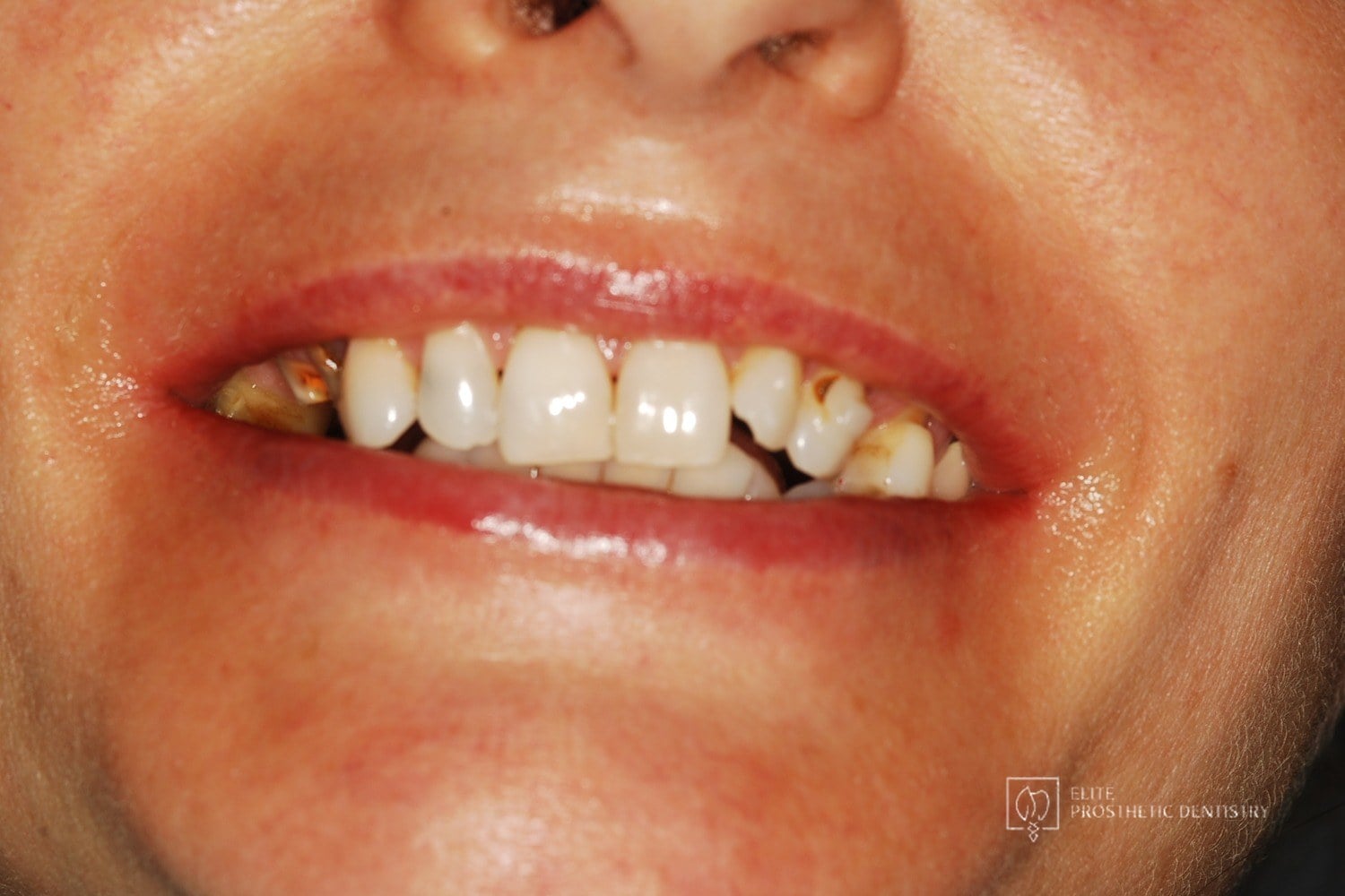 Before
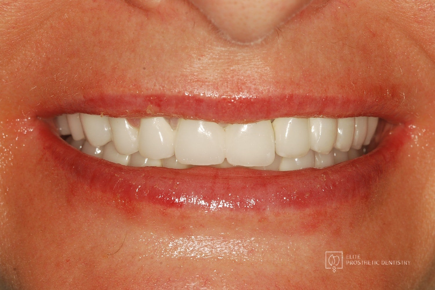 After
How Aging, Failing Dentistry Was Replaced with a Coordinated Full-Mouth Implant and Crown Reconstruction
The patient presented with throbbing upper-molar pain, long-standing infections, multiple root canals and unfinished prior treatment, generalized sensitivity, and several hopeless teeth.
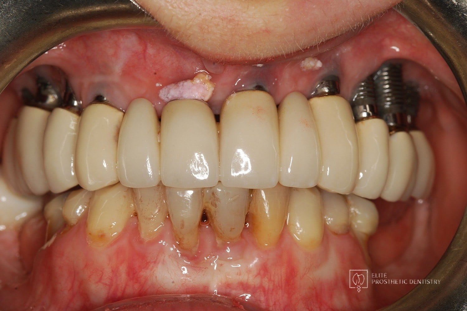 Before
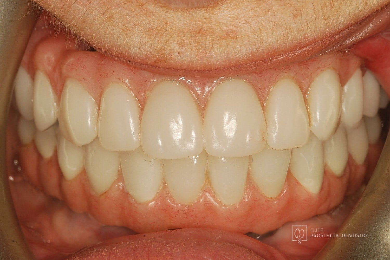 After
How Failing Implants and Severe Bone Loss Were Rebuilt Into a Stable Full-Arch Implant Reconstruction
The patient presented with significant complications from previously placed implants, severe bone loss, failing posterior implants, and an unesthetic full-arch bridge. Both arches required full reconstruction, with the upper arch showing the most advanced bone loss and instability.
 Before
 After
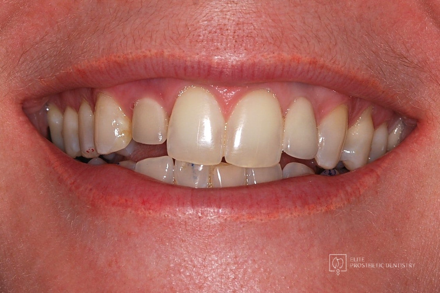
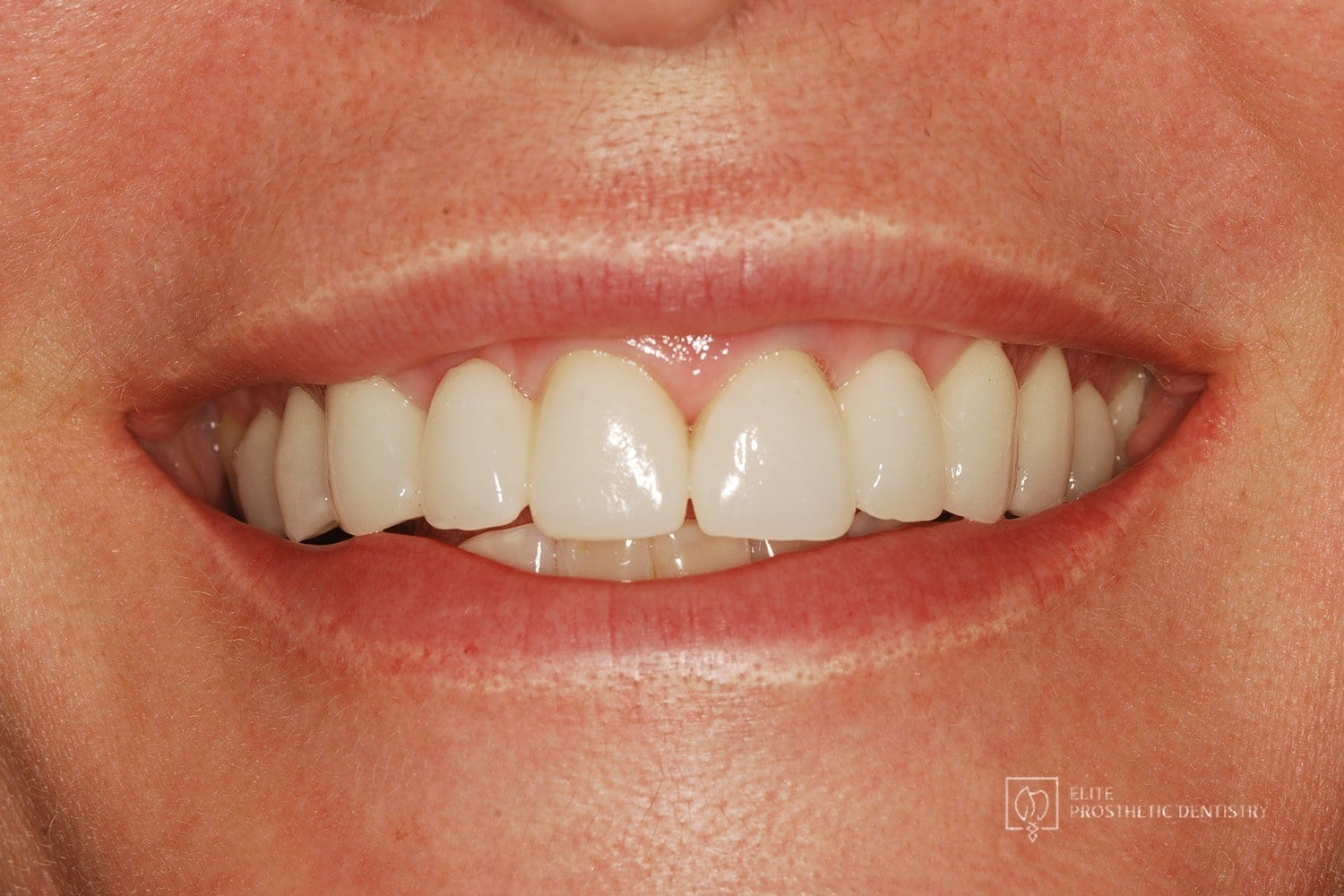
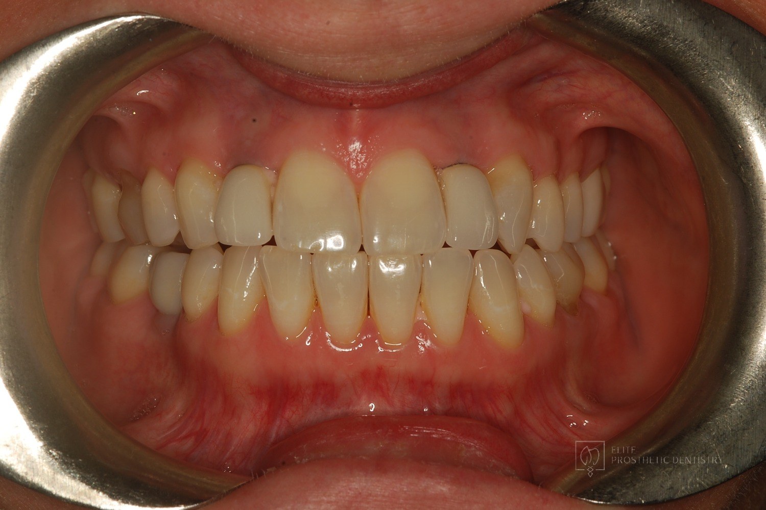
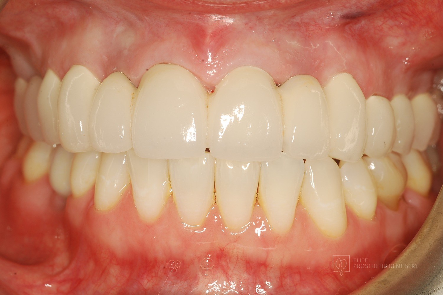
How Failing Lateral Incisor Implants Were Replaced with Custom Zirconia Bridges After Severe Bone Loss Made Replacement Implants Impossible
Comprehensive examination including CBCT imaging revealed significant bone loss surrounding implants that were placed outside the buccal plate, most likely due to placement without a CBCT-generated surgical guide. The implants were determined non-salvageable, and the alveolar ridge was too narrow to support replacement implants without extensive reconstruction.
 Before
 After
How Older Implant Crowns Were Redesigned for a Better Bite and More Natural Appearance
The patient came in after years of living with implant-supported crowns placed more than twenty years earlier that no longer looked or functioned well. CBCT evaluation, reviewed with a radiologist colleague, showed the implants had been placed too far to the buccal in very thin bone and could not support a healthy long-term restoration.
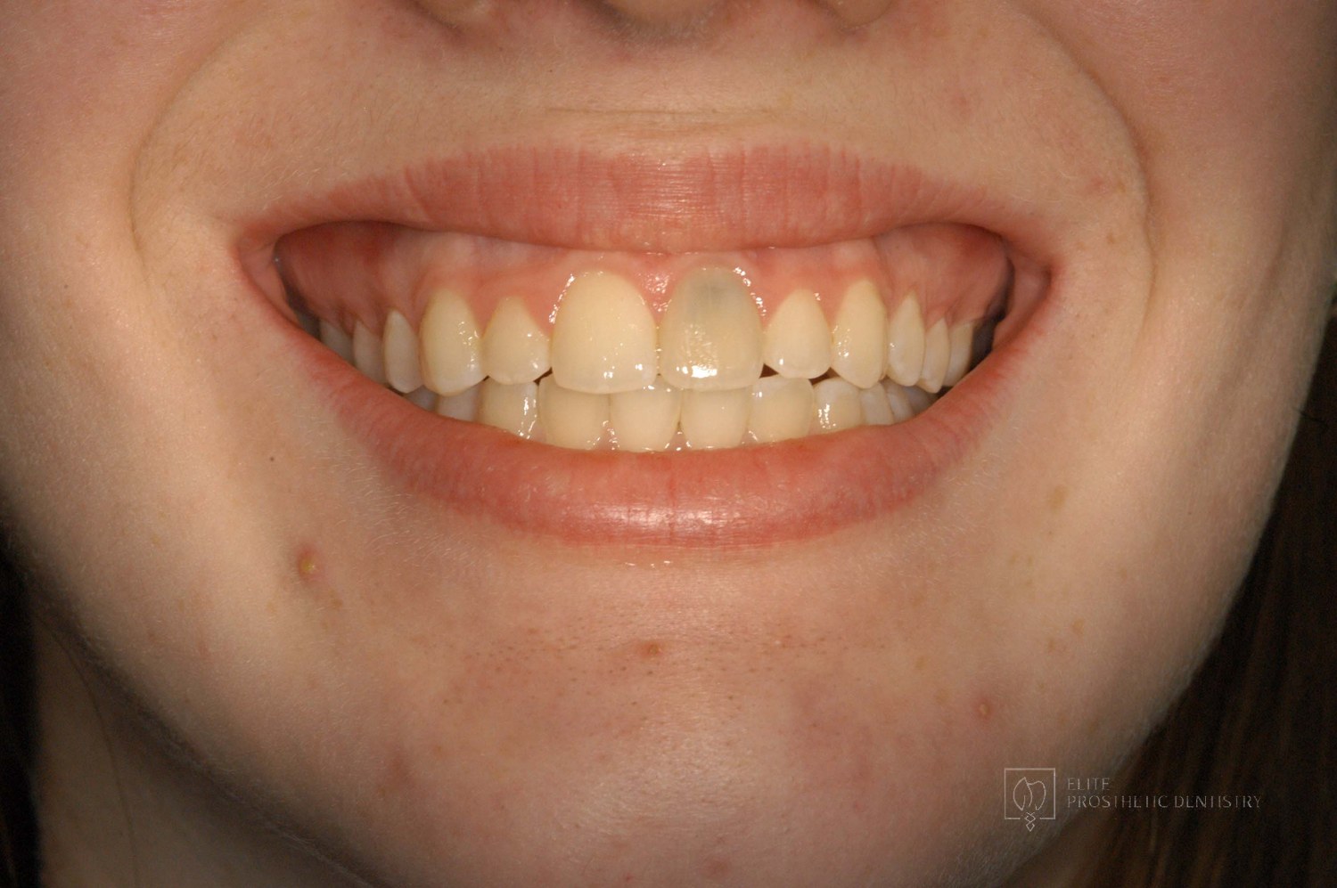 Before
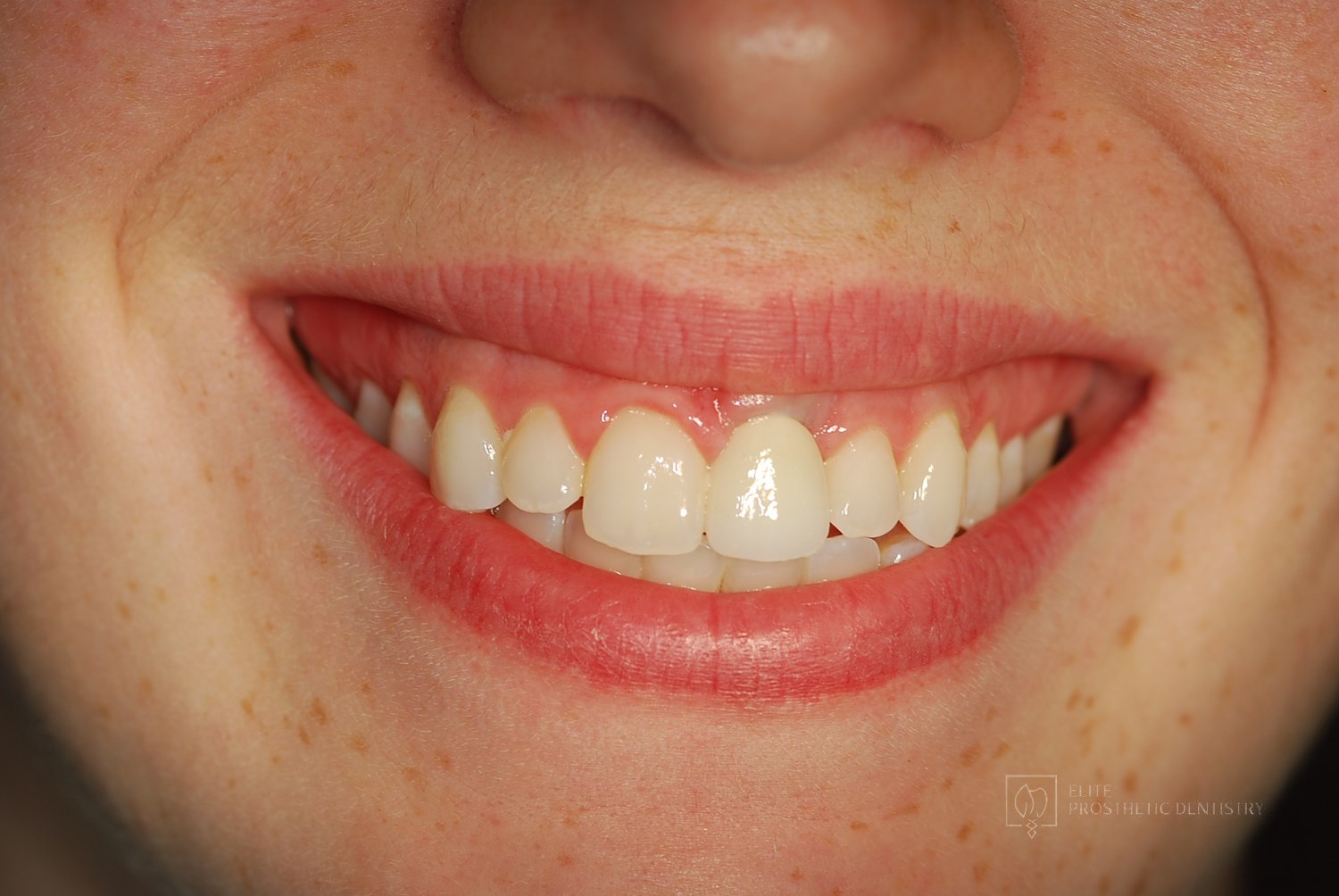 After
How a Front Tooth Lost to Childhood Trauma Was Rebuilt with Bone Grafting and a Long-Lasting Implant
A teenager was referred by her father after earlier trauma left her upper left front tooth slowly failing from root resorption. She was still growing, so an immediate implant was the wrong move. The tooth had to be maintained to buy time, then replaced correctly once she reached skeletal maturity.
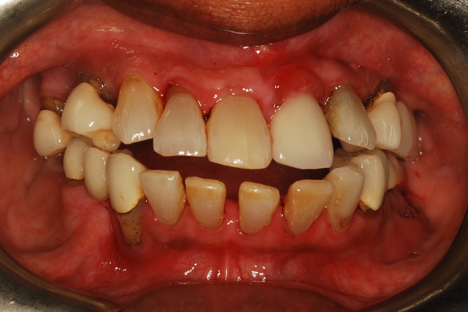 Before
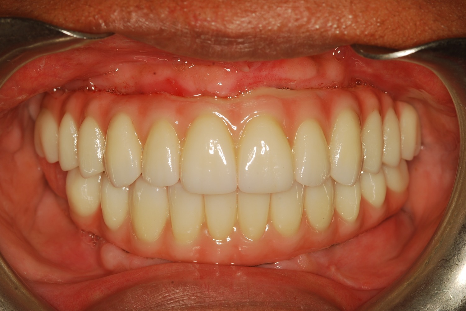 After
How Severe Bone Loss and Bite Dysfunction Were Rebuilt with All-on-6 Implants and a Milled Zirconia Hybrid Prosthesis
The patient presented with severe bone loss, advanced periodontal disease, malocclusion, and a dysfunctional bite that required full-arch rebuilding.
Related Articles
Deepen your knowledge with additional insights on this topic.
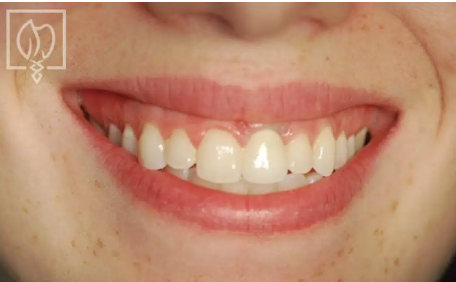 Dental Implants
If a Single Front Tooth Is Replaced with an Implant, Can It Look Natural?
Yes. See the four steps, with real case photos, that make a single front tooth implant indistinguishable from the natural tooth beside it. Washington, DC.
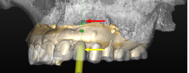 Dental Implants
What Is Precision Implant Placement (PIP)?
Precision Implant Placement plans each implant virtually on a CBCT scan, then delivers it with a custom surgical guide. See the three steps with real images.
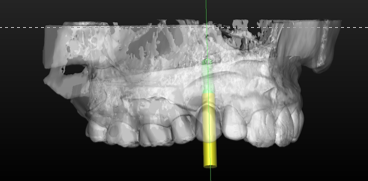 Dental Implants
What Is the Ideal Surgical Guide for Precision Implant Placement?
Not all surgical guides are equal. The gold standard is CBCT-based: planned virtually in 3D, 3D printed, and seated on your teeth. A DC prosthodontist explains.
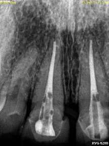 Dental Implants
When Should an Implant Not Be Done?
Healthy roots, thin bone, steep bony angles: a DC prosthodontist shows a real case where refusing implants was the right call, and what was done instead.
 Dental Implants
What Is Staged Implant Therapy?
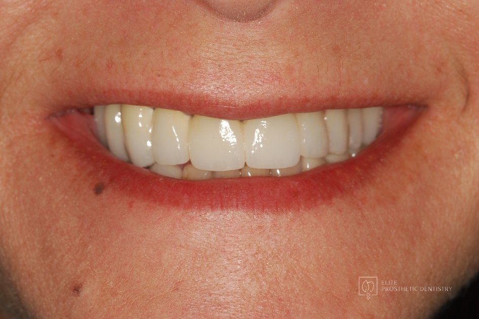
Staged implant therapy replaces failing teeth in phases while you keep fixed teeth the entire time. A DC prosthodontist walks through a real case in stages.
 Dental Implants
The Four Types of Dental Implants: Which One Is Right for You?
Endosteal, subperiosteal, zygomatic, and mini implants each solve a different problem. A DC prosthodontist explains the four types and who each one fits.
Have a patient with complex restorative, implant, bite, or esthetic needs?
Elite Prosthetic Dentistry provides specialist prosthodontic evaluation, treatment planning, and collaborative care while preserving the patient's relationship with the referring office. How referrals work here.
Ready to Transform Your Smile?
With 40+ years of experience and 3,900+ dental implants placed, Dr. Marlin and the team deliver dentistry planned to last. Request a specialist consultation.