

Why Am I Suddenly Getting Decay Under My Crowns? Dry Mouth, Recession and Aging Dentistry After 50
New decay under crowns that served for decades usually has a cause: dry mouth from medications, gum recession exposing margins, washed-out cement, a changed bite or diet. What to check, what it means for the teeth, and how to plan replacement.
Practice clinician: Gerald M. Marlin, DMD, MSD, prosthodontist, Elite Prosthetic Dentistry, Friendship Heights, Washington, DC. Last updated .
A patient in their fifties or sixties who has had crowns for twenty years without trouble, and is now being told at each visit that another one has decay underneath, is seeing a pattern rather than bad luck. Crowns do not decay; the teeth under them do, and when several start at once the conditions in the mouth have changed. This guide explains the usual causes, why they cluster after fifty, how decay under a crown is found, what it means for the tooth, and how to plan replacement so that the new crowns do not meet the same fate.
Where Decay Under a Crown Starts
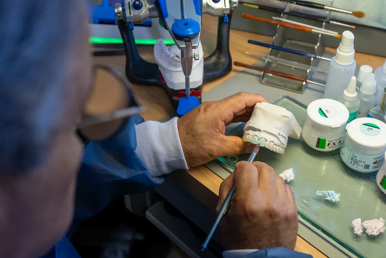
A crown covers the tooth above the gum line, and its edge, the margin, sits at or just below the gum. The margin is the weak point. It is a seam between two materials, sealed with cement, and it is the hardest part of the tooth to clean. Bacteria that collect there produce acid; saliva normally washes and neutralizes it. When the seam opens, the cement dissolves, the gum recedes to expose the seam, or the saliva is no longer doing its job, decay begins at the margin and spreads under the crown where no one can see it. On a tooth that has had a root canal, there is no nerve to signal a problem, and the first sign may be a crown that comes off with a soft tooth inside it.
Why It Clusters After Fifty
Dry mouth from medications. This is the single most common reason a stable mouth starts developing decay in middle age. Hundreds of medications reduce saliva: many blood pressure drugs, antidepressants, anti-anxiety medications, antihistamines, bladder medications, some pain medications. Patients who start one or two in their fifties, and a few more in their sixties, notice the dryness but rarely connect it to the new decay. Less saliva means more acid, more bacteria at the gum line, and decay at exactly the places crowns are vulnerable. Medical conditions and treatments can have the same effect.
Gum recession. Gums recede with age, with brushing habits, and with past gum disease. A crown margin placed under the gum in 1998 may now be exposed, with the root surface below it uncovered. Root surface is softer than enamel and decays more easily, and the exposed seam is harder to keep clean.
Cement wash-out. Older cements dissolve slowly over decades. A crown can be firmly in place while its margin has opened enough for bacteria to enter.
A changed bite. Wear, lost or shortened back teeth, and shifting over the years change how force reaches each crown. A margin that is flexed at every bite opens sooner. Clenching and grinding accelerate this. Our guide on why crowns keep breaking covers the bite side of the problem.
Diet and habits. More frequent snacking, sipping sweetened or acidic drinks through the day, lozenges for the dry mouth itself (many are sugared), and reduced dexterity for cleaning all raise the risk. None of these is a moral failing; they are conditions to be identified and managed.
Less frequent examination. Decay under a crown is found on X-rays and by probing. Patients who have gone years between examinations, or whose examinations did not include imaging of crowned teeth, lose the chance to catch it early.
How It Is Found, and What It Means for the Tooth
Decay under a crown is diagnosed with bitewing X-rays, careful probing of every margin, and, where the picture is unclear, a CBCT scan. It is not reliably visible to the eye, and it rarely hurts until late.
What happens next depends on how far it has gone:
- Early, at the margin: the crown is removed, the decay is cleaned out, the tooth is rebuilt and a new crown is made with the margin placed and sealed for the tooth’s current condition.
- Deeper, approaching the nerve: root canal treatment may be needed, then a post and core, then a new crown.
- Below the bone, or with too little tooth left: the tooth may not be savable, and the decision becomes implant, bridge or another design. Our guide on saving a tooth versus replacing it with an implant explains where that line is drawn, tooth by tooth.
The difference between the first group and the third is time. That is the argument for finding the cause and examining the other crowns now, rather than replacing them one at a time as each one fails.
Replacing Crowns Into the Same Conditions Does Not Work
A new crown placed into a dry, receded, acidic mouth with an uneven bite will develop the same problem, sometimes faster. A replacement plan for a patient with this pattern has two parts:
- Change the conditions. Identify the dry-mouth medications with the physician (sometimes an alternative exists; often the answer is management rather than change); saliva substitutes, prescription-strength fluoride, sugar-free products, cleaning aids for exposed margins, and an examination interval matched to the risk. Correct the bite where it is loading crowns unevenly, and protect against grinding.
- Design the new restorations for the conditions. Margin position and material chosen for an exposed root surface and a dry mouth, crowns designed for the bite as it is now, and, where several crowns are involved, one design for all of them rather than a series of matches to aging neighbors. The practice’s on-site laboratory allows that design to be refined with the patient present.
Our guide on when to replace old crowns and bridges covers the replace-now, plan-to-replace, or monitor judgment for each crown.
How Elite Prosthetic Dentistry Approaches This Pattern
Dr. Gerald M. Marlin treats recurrent decay under crowns as a mouth-level problem with a cause, not as a series of unrelated crowns. The evaluation images and probes every existing restoration, records the medications and the saliva situation, assesses the bite, and classifies each crown as replace now, plan to replace, or monitor. Where teeth are savable, they are saved; where a few are not, the plan addresses them alongside the rest. The practice sees patients for both comprehensive reconstruction and proactive replacement of aging dentistry, and this pattern is the most common reason a patient moves from the second category into a larger plan than they expected.
When Another Setting Fits Better
One crown with decay in a mouth whose other restorations are sound and whose saliva and bite are normal is a routine replacement for a general dentist. Dry mouth that is severe or accompanied by other symptoms may warrant a physician’s evaluation for an underlying condition. Patients with active periodontal disease across the mouth need that treated before restorative planning.
Documented Cases
- Loose, failing crowns, recurrent decay under restorations, abscesses and significant dry mouth: strategic teeth saved and both arches rebuilt.
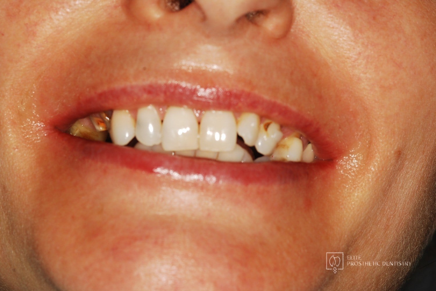
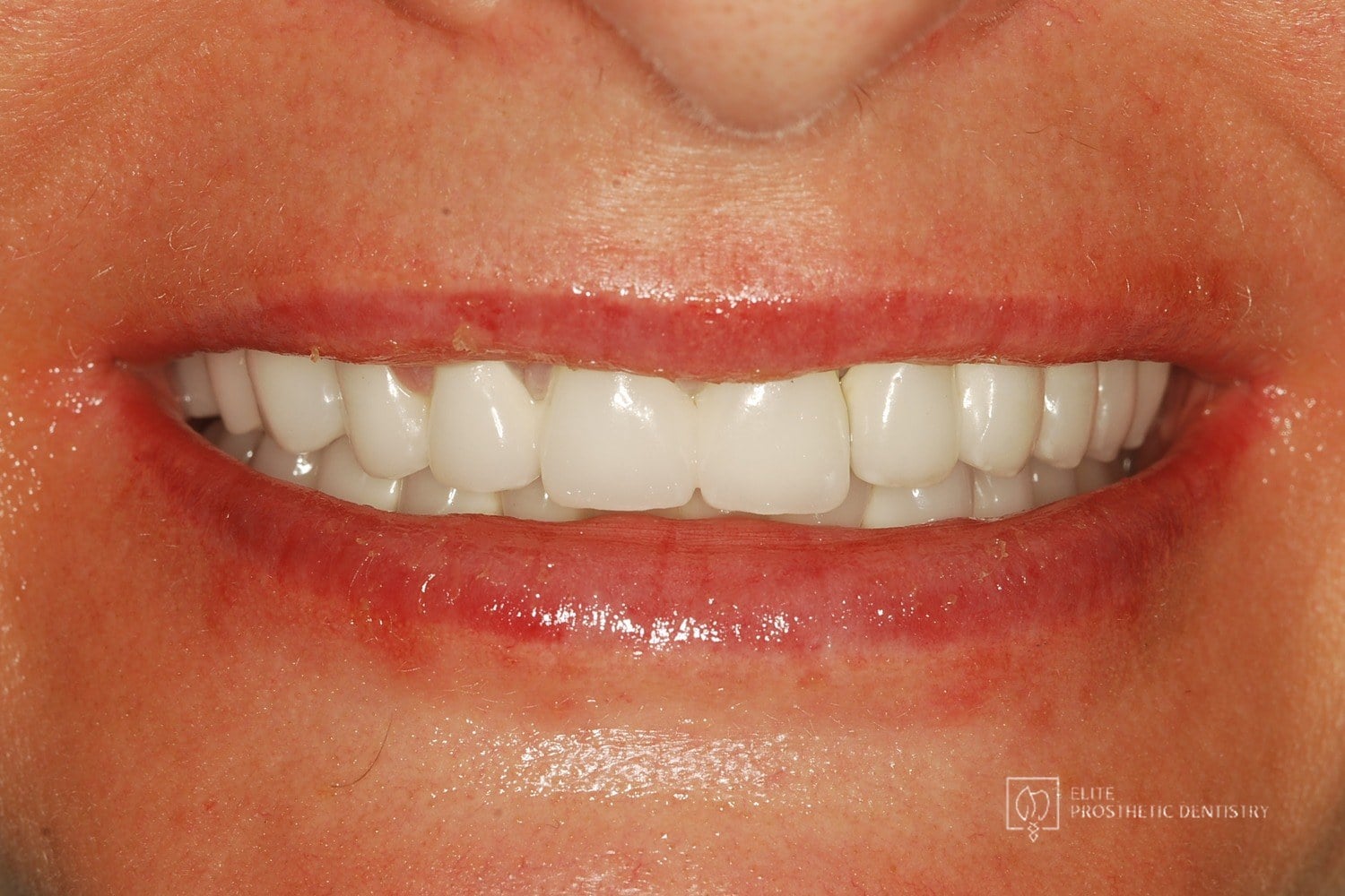
- Recurrent decay beneath mismatched crowns treated with root canals and buildups before a customized bridge.
- Aging, opaque restorations replaced with customized ceramic restorations.
- Aging crowns and a long-standing bridge rebuilt with a coordinated restorative plan.
More cases are organized in the replacing aging dentistry success story hub.
Related Decision Guides
- When should old crowns and bridges be replaced?
- Should I save the tooth or replace it with an implant?
- Why do my crowns keep breaking?
- General dentist vs. prosthodontist for aging crowns, bridges and implant restorations
- Signs your dental work is wearing out
Request an Evaluation of Your Crowns
If decay has been found under one crown and you have others of the same age, request an appointment or call (202) 244-2101. Bring your medication list and recent X-rays; the evaluation will look for the cause and examine the rest.
Your Best Smile Is Within Reach
Request a specialist consultation with Dr. Marlin to discuss your situation and the most appropriate path forward.
Frequently Asked Questions
Can a tooth decay under a crown?
Yes. The crown itself cannot decay, but the tooth beneath it can, and the usual entry point is the margin where the crown meets the tooth. Decay there is painless until it is advanced, because the crown covers the tooth and the nerve may already have been treated. It is found on X-rays and by probing the margin, which is why examinations with imaging matter more as crowns age.
Why would this start now, after decades without a problem?
Something changed. The most common changes after fifty are reduced saliva from medications or medical conditions, gum recession that exposes a margin that was once protected, cement that has slowly washed out at an edge, a bite that has shifted and now loads the crown differently, or a diet or habit that has changed. The crowns did not become worse; the conditions they live in did. Finding the cause matters, because new crowns placed into the same conditions will fail the same way.
Which medications cause dry mouth?
Hundreds do. Common groups include blood pressure medications, antidepressants and anti-anxiety medications, antihistamines, bladder medications, some pain medications and many others; taking several together compounds the effect. Saliva neutralizes acid and clears bacteria, so less of it means more decay, often at the gum line and at crown margins. If you have started new medications in recent years and are seeing new decay, the two are probably connected.
If decay is found under a crown, does the tooth have to be extracted?
Usually not, if it is found early. The crown is removed, the decay is cleaned out, the tooth is rebuilt as needed and a new crown is made. If the decay has reached the nerve, root canal treatment may be needed first. If it has gone below the bone or left too little tooth, the tooth may not be savable. Our guide on saving a tooth versus replacing it explains how that line is drawn.
Should I replace all my old crowns before they decay?
No. Crowns with intact margins, no decay on imaging, healthy gum and a stable bite can be monitored. Replacement is planned for crowns with open or exposed margins, decay, fracture or a changed bite, and the plan is made for the mouth as a whole rather than one crown at a time. Our guide on when to replace old crowns and bridges covers the judgment.
Related Patient Success Stories
Explore similar patient success stories demonstrating our expertise in advanced prosthetic dentistry.
 Before
 After
How Aging, Failing Dentistry Was Replaced with a Coordinated Full-Mouth Implant and Crown Reconstruction
The patient presented with throbbing upper-molar pain, long-standing infections, multiple root canals and unfinished prior treatment, generalized sensitivity, and several hopeless teeth.
 Before
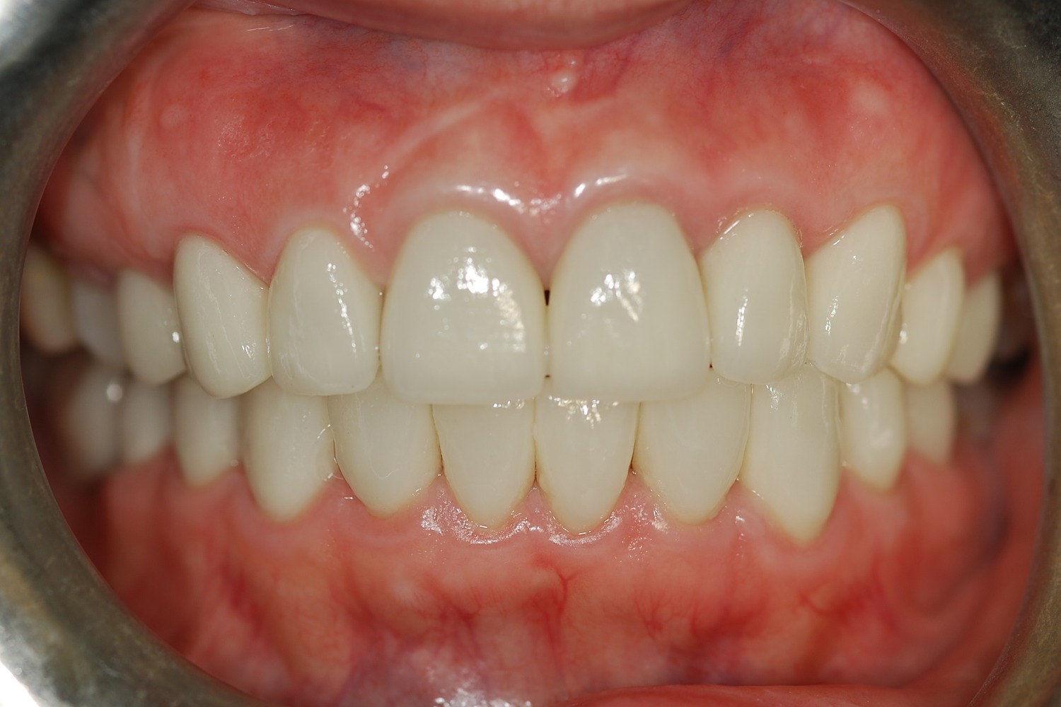 After
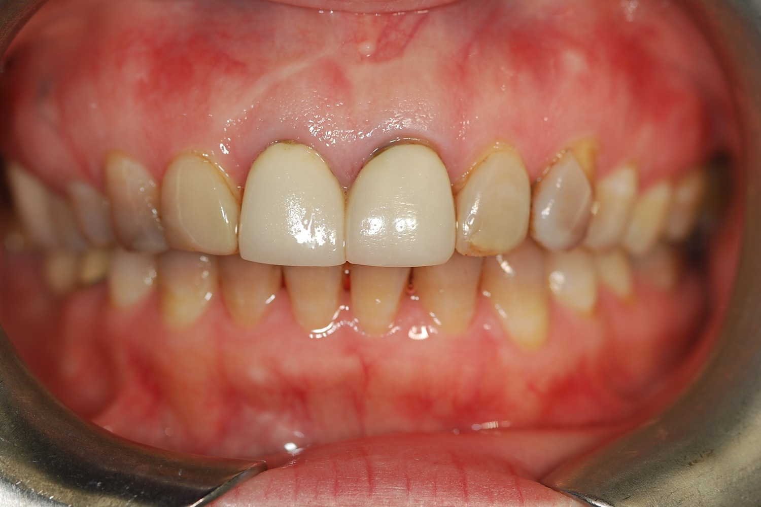
How Aging, Opaque Restorations Were Replaced with Customized Ceramic Restorations Designed for Long-Term Natural Esthetics
The existing restorations appeared opaque, worn, and unnatural over time, affecting both confidence and overall smile harmony.
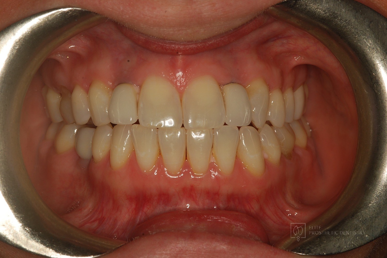 Before
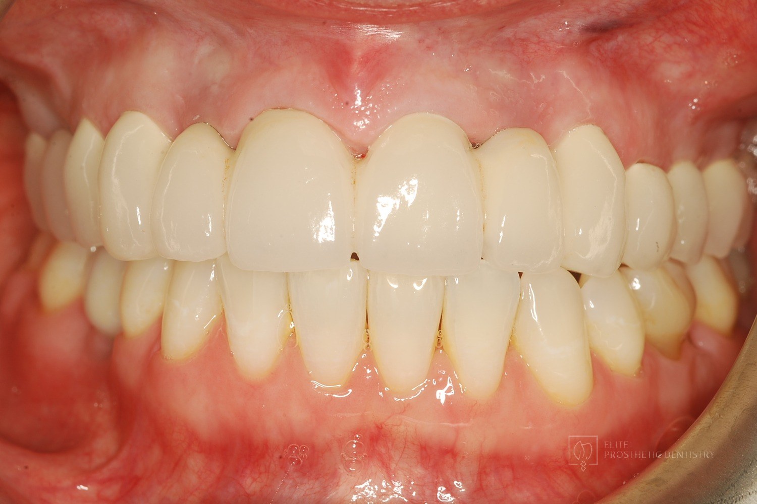 After
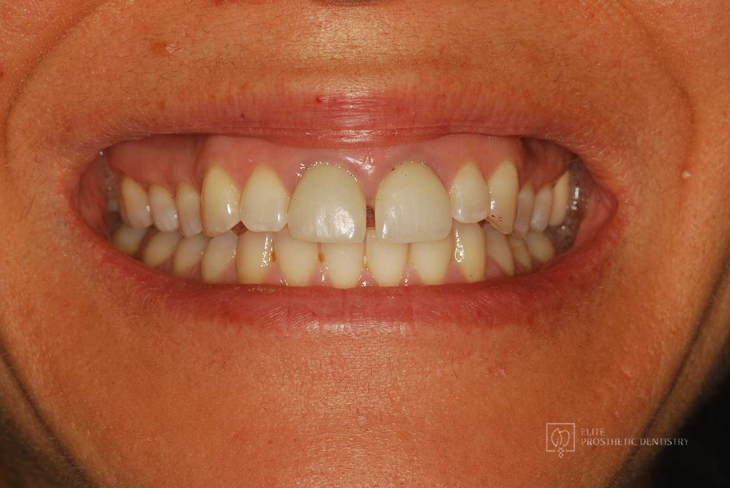
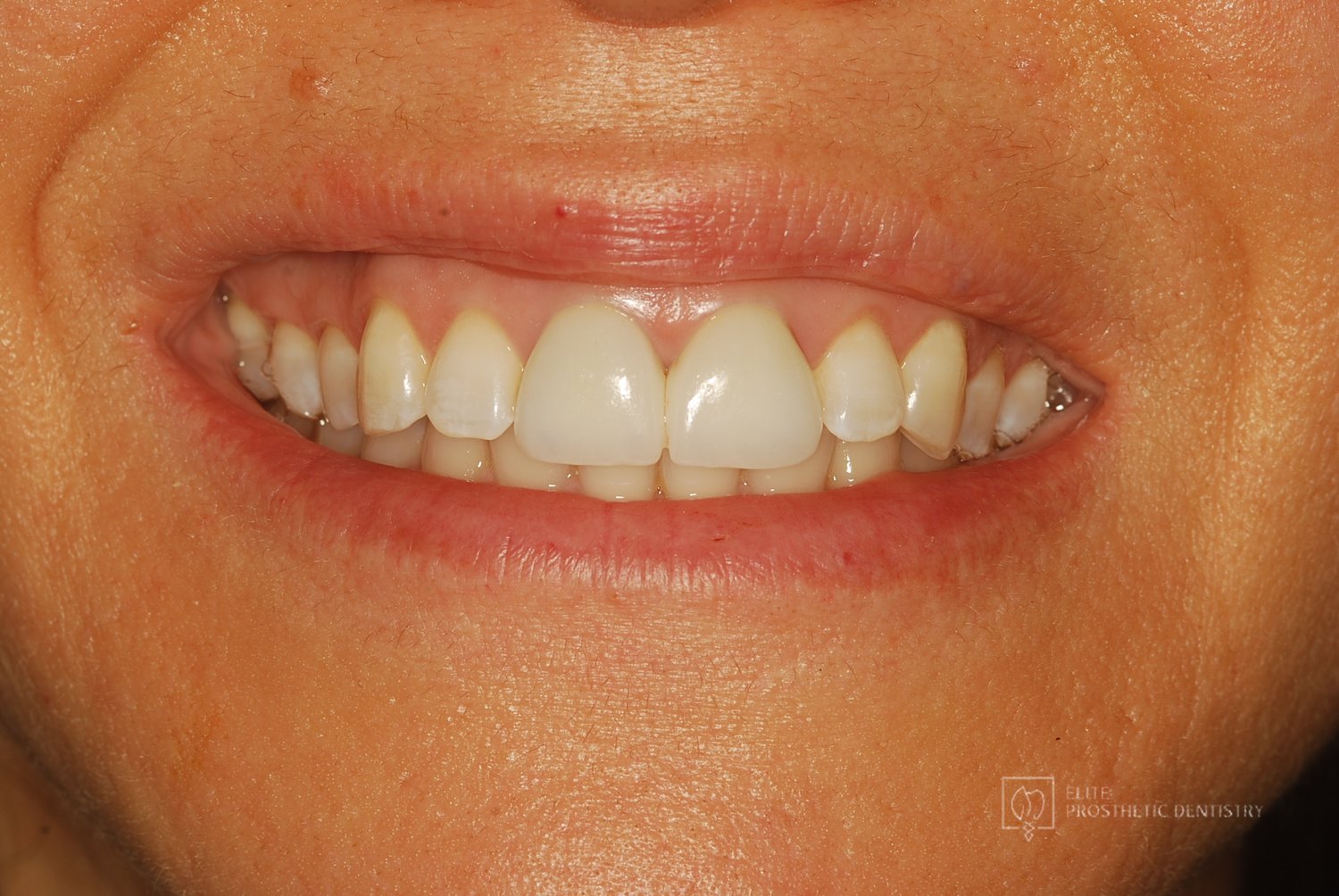
How Older Implant Crowns Were Redesigned for a Better Bite and More Natural Appearance
The patient came in after years of living with implant-supported crowns placed more than twenty years earlier that no longer looked or functioned well. CBCT evaluation, reviewed with a radiologist colleague, showed the implants had been placed too far to the buccal in very thin bone and could not support a healthy long-term restoration.
 Before
 After
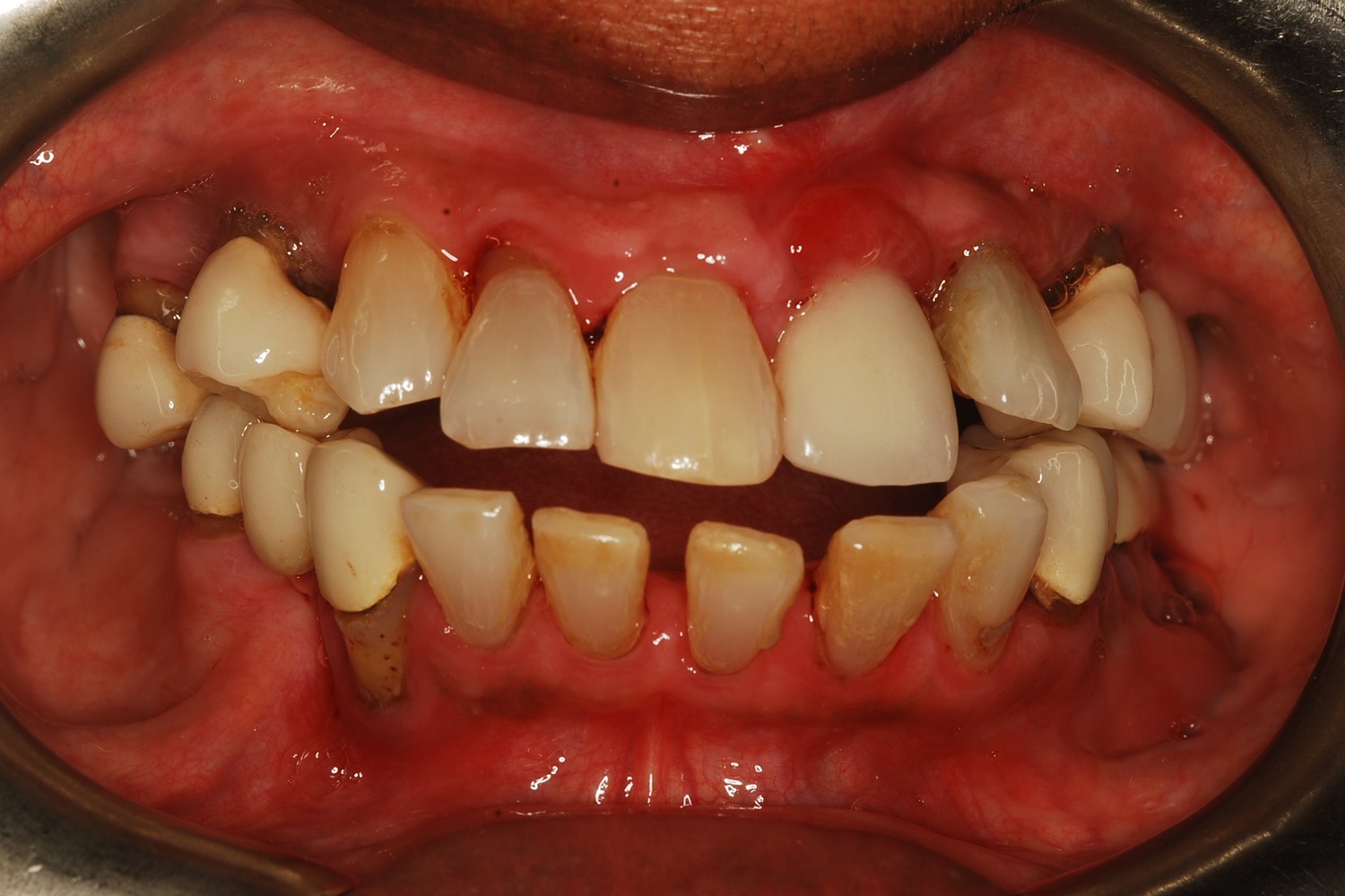
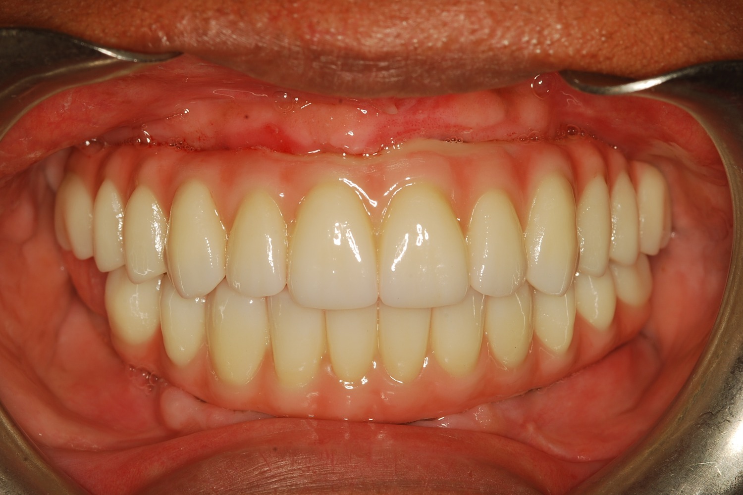
Two Front Teeth Saved From Extraction: A Second Opinion, Custom Gold Posts, and Crowns Made to Last
Two upper central incisors with failed root canal treatment and recurrent decay had been recommended for extraction and implant replacement. A CBCT evaluation showed that removing the roots from their thin facial bone housing could create a visible esthetic defect in the gum and bone contour, made worse by the patient's high lip line.
 Before
 After
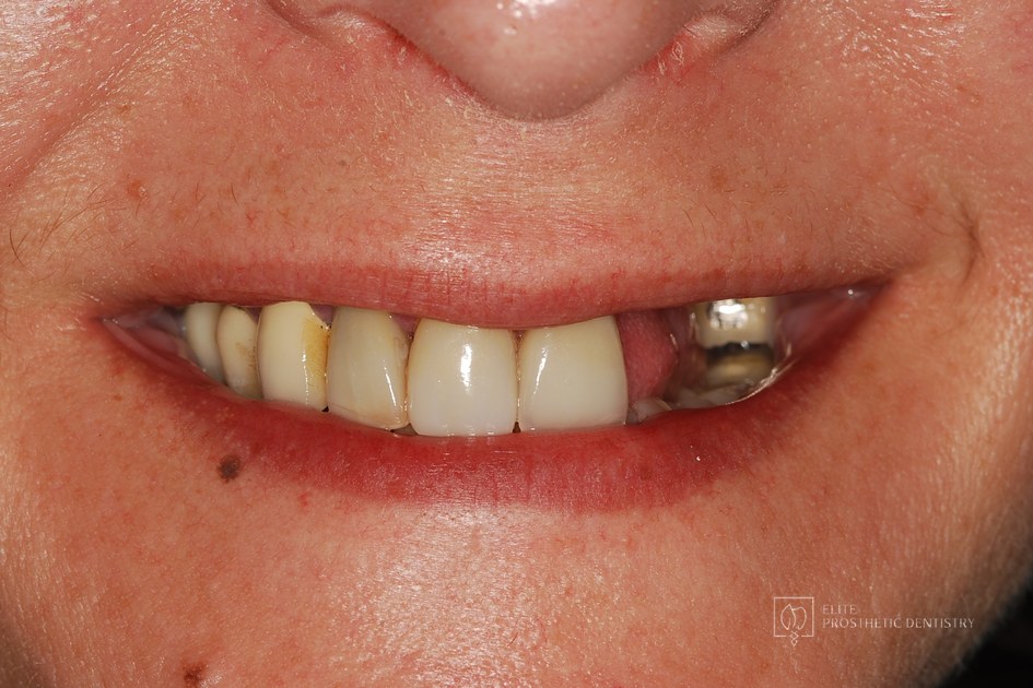
How Severe Bone Loss and Bite Dysfunction Were Rebuilt with All-on-6 Implants and a Milled Zirconia Hybrid Prosthesis
The patient presented with severe bone loss, advanced periodontal disease, malocclusion, and a dysfunctional bite that required full-arch rebuilding.
 Before
 After
Implant Supported Reconstruction: Failing Bridgework and Missing Back Teeth Rebuilt with Coordinated Specialist Care
Referred by another dental specialist with severe bone resorption on the upper left, multiple broken-down lower teeth requiring extraction, and failing lower back teeth that had left the bite without solid support. No single procedure, and no provider working in isolation, could rebuild a situation this interconnected.
Related Services
Ready to Transform Your Smile?
With 40+ years of experience and 3,900+ dental implants placed, Dr. Marlin and the team deliver dentistry planned to last. Request a specialist consultation.