
Multi-Faceted Treatment for a Patient Unhappy With the Appearance of Her Crowns, Teeth, and Gums
Documented before-and-after view
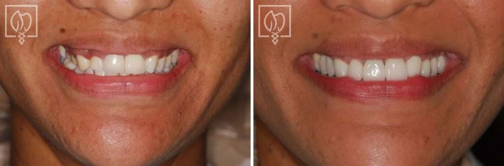
A multi-faceted smile restoration built around one coordinated plan
Coordinated smile restoration in Washington, DC. This documented case at Elite Prosthetic Dentistry combined orthodontic preparation, a single dental implant, new front-tooth crowns, and minor gum contouring for a patient who was unhappy with how her teeth and gums worked together as a smile. Treatment was planned by Dr. Gerald Marlin, D.M.D., M.S.D., a prosthodontist focused on complex restorative and esthetic dentistry, and carried out step by step with his team.
Case at a Glance
- Treatment
- Orthodontic repositioning, an implant with custom crown, new front-tooth crowns, and gingival contouring
- Approach
- One sequenced plan: reposition the teeth, replace the missing tooth, restore, then balance the gum line
- Case Type
- Replacing aging dentistry / Cosmetic-functional correction
- Patient Concern
- Unhappy with how her front-tooth crowns, a missing tooth occupied by the wrong-sized tooth, and an uneven gum line all affected the look of her smile.
- Decision Point
- Restore the front crowns as they were, or move the misplaced canine into proper position first so the implant and crowns could be built around a corrected arrangement.
- Outcome
- The coordinated sequence produced a balanced, completely natural-looking smile, with the repositioned teeth, implant, new crowns, and evened gum line all supporting each other.
- Why It Matters
- Shows that interconnected esthetic problems, tooth position, gum architecture, and restorations, cannot be solved separately, and that planning the final arrangement first and sequencing each discipline produces a natural result.
The presenting condition
Many patients arrive at the practice unhappy with the appearance of their smile. What set this case apart was that the dissatisfaction did not trace to a single cause. The patient was very unhappy with how her teeth and gums together were affecting her smile, and with it her appearance and confidence. During her consultation, Dr. Marlin identified three distinct problems that would each need to be addressed for the result to look natural.
First, the crowns on her two front teeth no longer served her esthetically. They were wide and square in form and made of an opaque white material that contrasted with her gingiva and complexion, and they did not match the neighboring natural teeth in color, translucency, or shape, so they stood out as restorations rather than blending in as teeth.
Second, she was missing a lateral incisor, one of the narrower teeth that normally sits beside the front teeth. Her left canine, a naturally larger tooth, had come to occupy the lateral incisor’s position, which threw off the proportions of the smile.
Third, her gum line was uneven. The tissue sat too low around the right canine and lateral incisor area, a gap showed between her two front teeth, and on the left side the tissue was positioned noticeably higher than on the right, leaving the smile visually unbalanced.
Clinical Findings
- →Front-tooth crowns that were wide and square, in an opaque shade that no longer blended with the adjacent natural teeth
- →Contrast between the opaque crown material and the patient's gingiva and complexion
- →A missing lateral incisor, with the larger left canine occupying its space
- →An uneven gingival line, lower around the right canine and lateral incisor area and higher on the left
- →A visible gap between the two front teeth
Why this case required prosthodontic-level planning
None of these three problems could be solved in isolation, and that is the defining feature of the case. New crowns made for the front teeth as they stood would still have sat beside a canine in the wrong position and under an uneven gum line, and the result would have looked improved rather than natural. The gum tissue frames every restoration. The tooth positions set the proportions the crowns must follow. And the crowns themselves succeed only when both of those are right. Prosthodontic planning works from the endpoint backward: define the final tooth arrangement, the gum frame, and the restorative design first, then sequence each discipline so that every step sets up the next.
The decision behind the result: reposition the teeth before restoring them
The pivotal judgment in this case was to begin with tooth movement rather than restoration. The canine occupying the lateral incisor space could not simply be disguised. A canine is a larger tooth by nature, and restoring the smile around it would have locked the wrong proportions into the final result. Orthodontic treatment came first, moving the canine out of the lateral incisor position and bringing the teeth into a more natural, balanced arrangement. Only then did the space exist to replace the missing lateral incisor properly with an implant, design front-tooth crowns to correct proportions, and finish the gum line around teeth that were finally in the right places. It is a slower path than restoration alone, and it is the reason the finished smile looks like it was never treated.
The treatment plan
-
1
Comprehensive consultation and diagnosis
Dr. Marlin identified the three interconnected problems, crown esthetics, tooth position, and gum balance, and mapped the complete endpoint before any treatment began.
-
2
Orthodontic repositioning
The larger canine was moved out of the space where the lateral incisor belonged, repositioning the teeth into a more natural and balanced arrangement.
-
3
Implant placement for the missing lateral incisor
With the space restored, a dental implant was placed with precision to provide optimal support for a custom crown matched to her natural teeth.
-
4
New crowns for the two front teeth
The opaque crowns were replaced with natural-looking crowns with appropriate translucency and internal coloration, carefully shade-matched and designed to reflect light the way natural teeth do. The replacement restorations were developed in direct coordination with the practice's in-house laboratory to keep control over shade, contour, and fit.
-
5
Gingival contouring
Minor gum surgery evened and reshaped the tissue line so the gums frame the teeth symmetrically across the entire smile.
The outcome
With treatment complete, the patient achieved a completely natural-looking smile, with precision-made crowns and restorations designed to provide both beauty and function for many years. Each element supports the others. The orthodontic phase set correct positions, the implant filled the missing-tooth space with independent support, the new crowns blend with the neighboring teeth instead of standing apart from them, and the balanced gum line frames it all evenly. The documentation for the case sums it up simply: a smile she could finally be proud of.
Result Highlights
- ✓Three interconnected problems resolved through one sequenced plan
- ✓Canine moved out of the lateral incisor space into a natural, balanced position
- ✓Missing lateral incisor replaced with a precisely positioned implant and custom crown
- ✓Front-tooth crowns remade with lifelike translucency and internal coloration
- ✓Gum line evened and reshaped to frame the smile symmetrically
- ✓A natural-looking result the patient could finally feel confident about
Who this case may sound familiar to
This case tends to resonate with patients in a few recognizable situations:
- You have crowns on your front teeth that looked fine once but no longer blend with the rest of your smile.
- You are missing a tooth, other teeth have shifted into the space, and the spacing has never looked quite right.
- Your gum line sits higher on one side than the other, and you notice it every time you smile.
- You have been offered a single fix, new crowns or gum reshaping alone, but suspect the problems are connected.
- You want the sequence planned once and done correctly rather than improvised one restoration at a time.
If any of those describe where you are, a consultation with Dr. Marlin can establish the diagnostic picture and the specific options for your case.
Frequently asked questions
Why do some crowns look opaque or flat next to natural teeth?
Natural enamel transmits light, so a natural tooth has depth, translucency, and subtle internal color variation. Crowns made from a more opaque material reflect light from the surface instead, which can make them look flat or noticeably whiter than the teeth beside them. Crowns designed with layered translucency and internal coloration allow light to behave the way it does in a natural tooth.
Can a missing lateral incisor be replaced with a dental implant?
Yes, when there is adequate space and bone. A lateral incisor is a narrow tooth, so the space often has to be prepared first. If a neighboring tooth has moved into the gap, orthodontic treatment re-opens the correct space before the implant is placed and restored with a custom crown.
Why would orthodontics come before an implant and new crowns?
Because an implant cannot be moved once it has integrated, and crowns are designed to fit the positions the teeth are in. Setting the final tooth arrangement first means the implant is placed in the right position and the new crowns are made to correct proportions rather than compensating for crowding or drift.
What does gum contouring change about a smile?
The gum line is the frame around the teeth. When tissue sits noticeably higher on one side or lower in one area, the smile can look unbalanced even when the teeth themselves are attractive. Minor gingival surgery reshapes and evens the tissue line so the teeth are framed symmetrically.
Why see a prosthodontist when several problems affect one smile?
A prosthodontist is trained to plan the smile as a connected system: tooth position, gum architecture, restorations, and bite together. When a case involves orthodontics, an implant, crowns, and gum surgery, that endpoint-first planning determines the sequence and keeps every discipline working toward the same final result.
More about the work behind this case
This case sits at the intersection of dental crowns, dental implants, and cosmetic dentistry. The endpoint-first sequencing and in-house laboratory control that shaped it are part of the practice philosophy behind every complex esthetic case at the practice.
Elite Prosthetic Dentistry treats patients from across the DMV including Bethesda, Chevy Chase, McLean, Arlington, Potomac, and Great Falls, with a record of out-of-area patients traveling to the practice for complex restorative care.
Related Services
Related Patient Success Stories
Explore similar patient success stories demonstrating our expertise in advanced prosthetic dentistry.
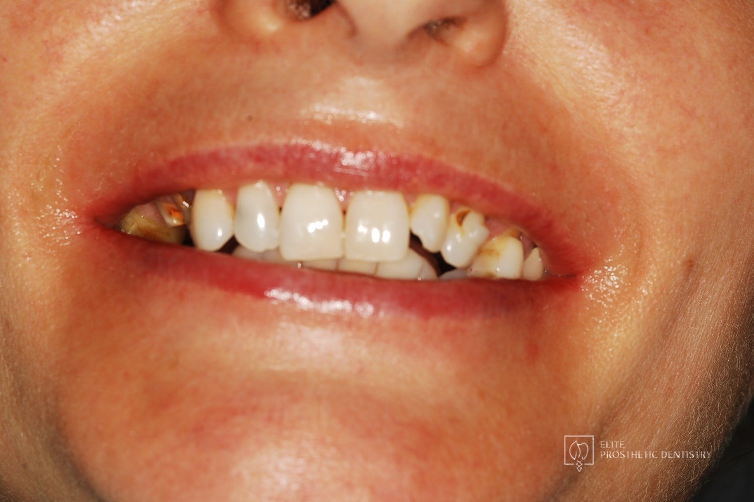 Before
 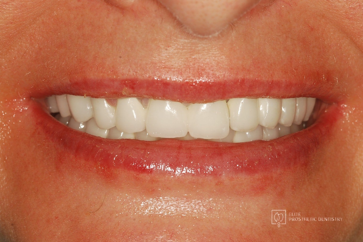 After
After How Aging, Failing Dentistry Was Replaced with a Coordinated Full-Mouth Implant and Crown Reconstruction
The patient presented with throbbing upper-molar pain, long-standing infections, multiple root canals and unfinished prior treatment, generalized sensitivity, and several hopeless teeth.
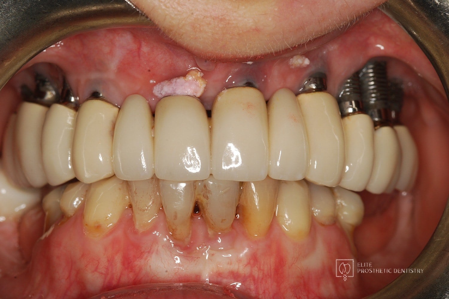 Before
 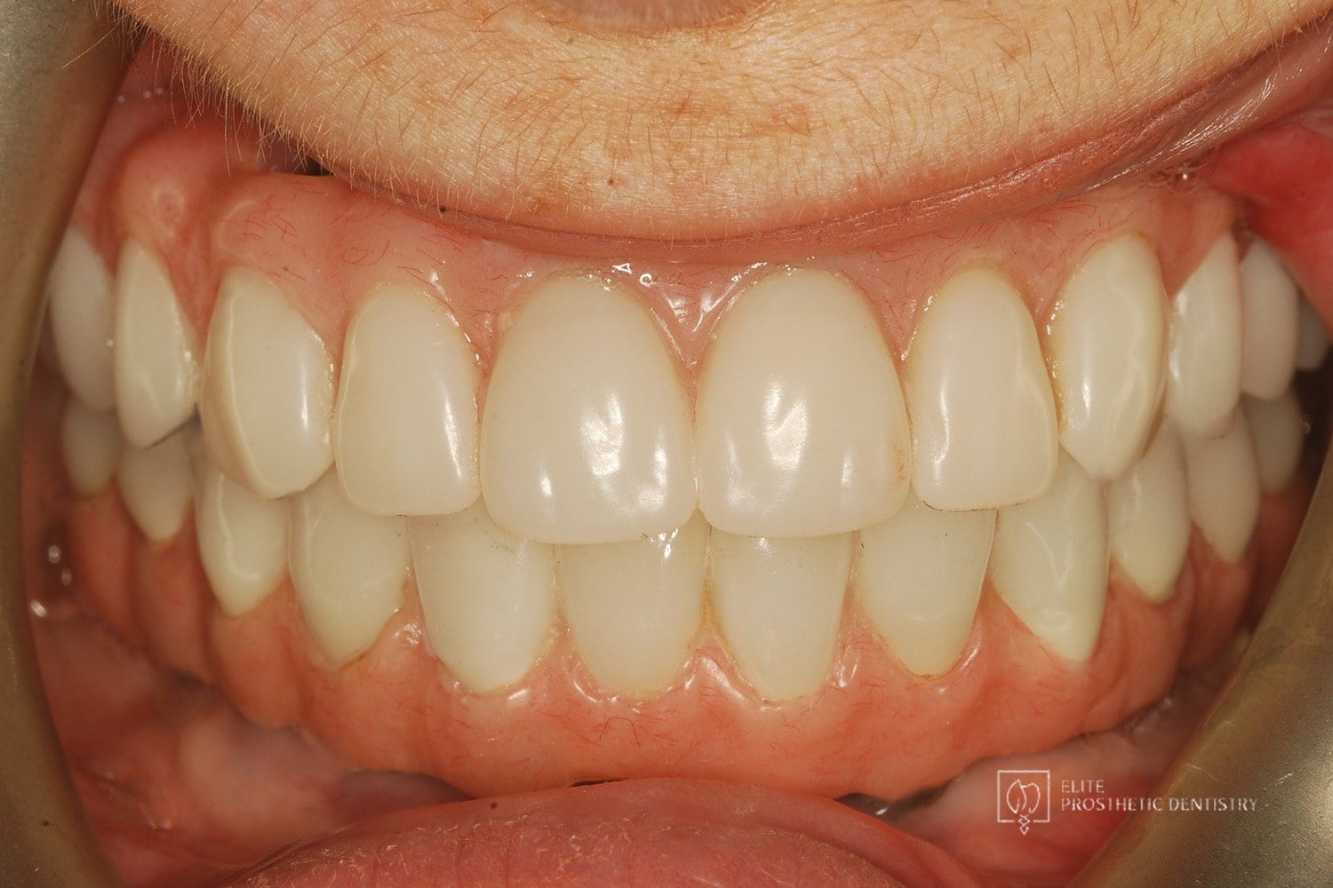 After
After How Failing Implants and Severe Bone Loss Were Rebuilt Into a Stable Full-Arch Implant Reconstruction
The patient presented with significant complications from previously placed implants, severe bone loss, failing posterior implants, and an unesthetic full-arch bridge. Both arches required full reconstruction, with the upper arch showing the most advanced bone loss and instability.
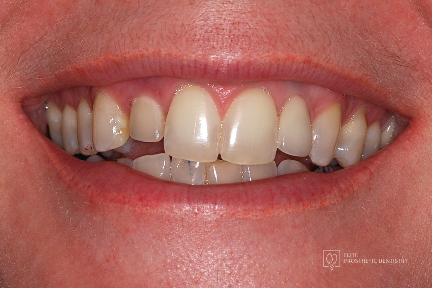 Before
 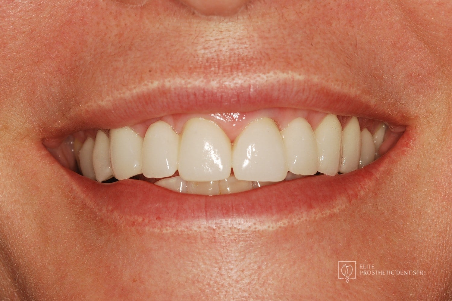 After
After How Failing Lateral Incisor Implants Were Replaced with Custom Zirconia Bridges After Severe Bone Loss Made Replacement Implants Impossible
Comprehensive examination including CBCT imaging revealed significant bone loss surrounding implants that were placed outside the buccal plate, most likely due to placement without a CBCT-generated surgical guide. The implants were determined non-salvageable, and the alveolar ridge was too narrow to support replacement implants without extensive reconstruction.