
Front Tooth Missing for Ten Years and Told There Was Not Enough Bone: Grafting, Ridge Augmentation, and a Guided Implant
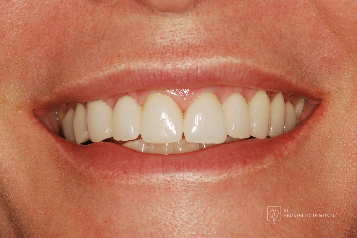
Smile view
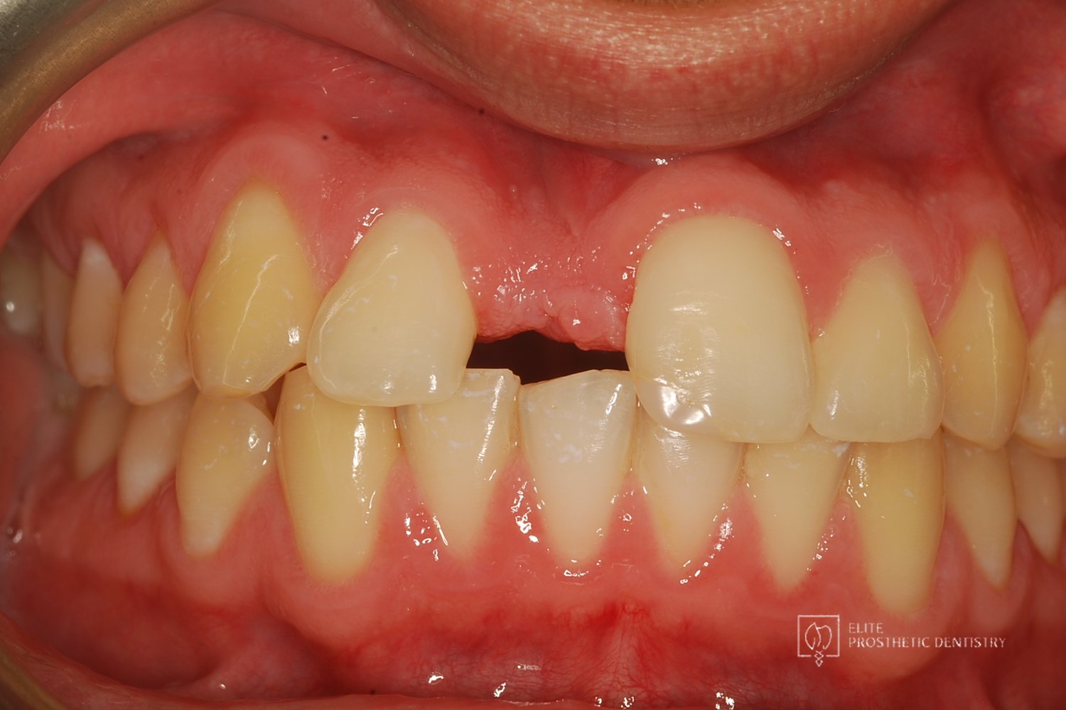 Before
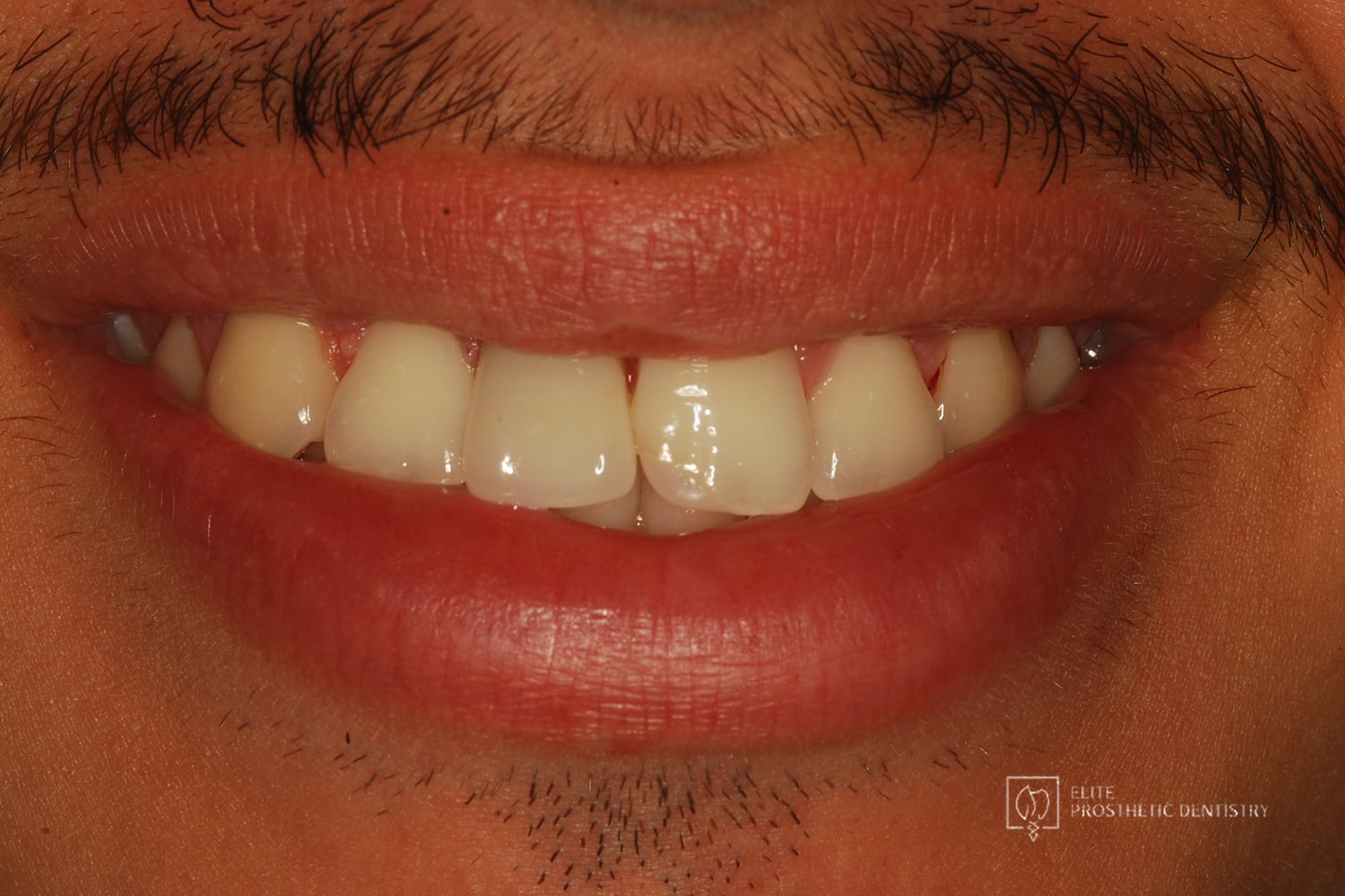 After
Completed result
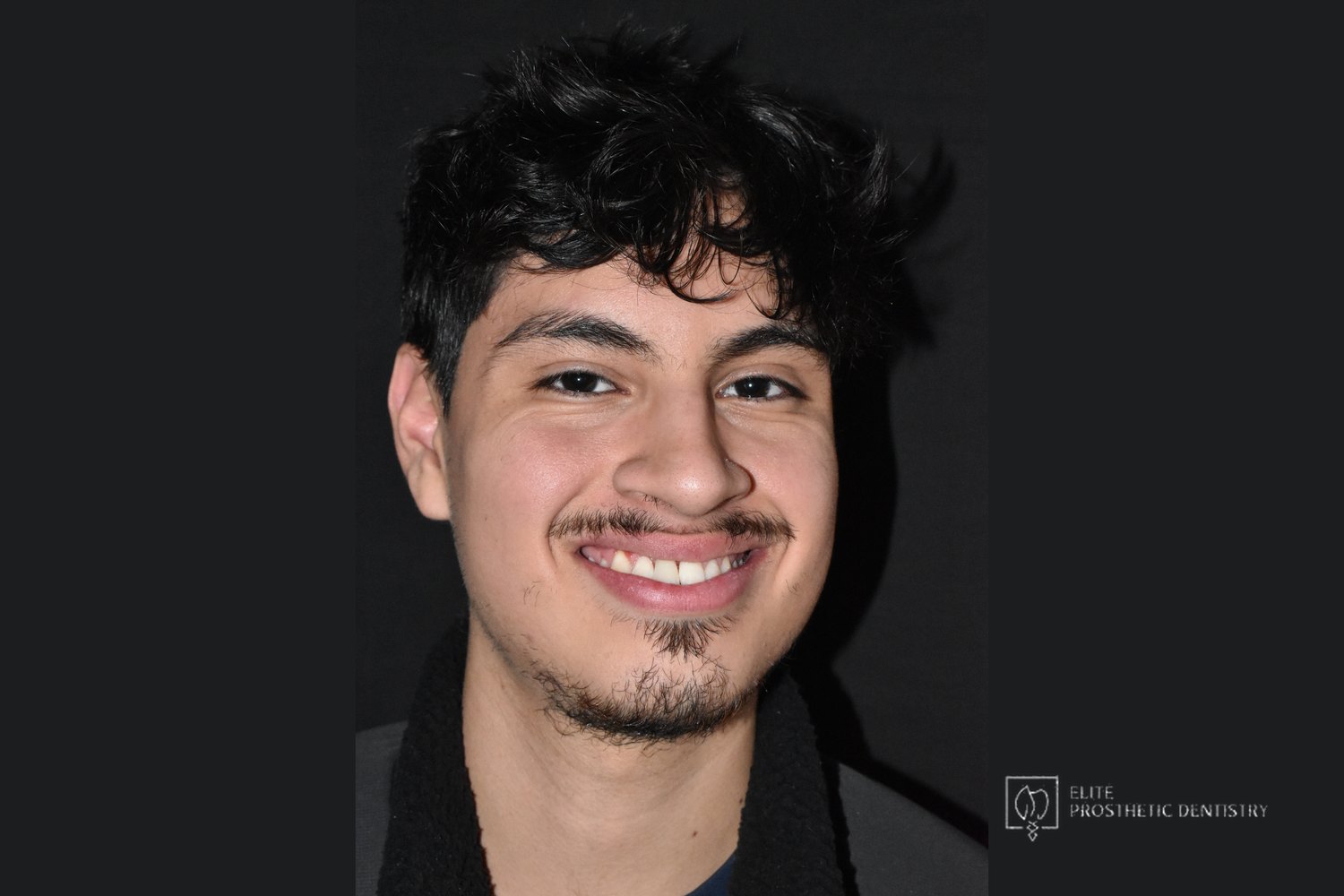
A front tooth missing since childhood, rebuilt after being told it was too late
Replacing an upper central incisor missing for more than ten years in Washington, DC for a 19-year-old who had lost the tooth to a facial injury as a child. This documented case at Elite Prosthetic Dentistry used CBCT evaluation, two bone grafts with ridge augmentation, and a custom surgical guide to place an implant in a site where the patient had been told elsewhere that not enough bone remained. Treatment was planned and completed by Gerald Marlin, D.M.D., M.S.D., a prosthodontist who plans bone reconstruction and the final crown together.
Case at a Glance
- Treatment
- Single-tooth implant replacement of an upper central incisor after severe long-term bone loss
- Approach
- CBCT evaluation of the defect, two bone grafts with ridge augmentation, guided implant placement for ideal position and angulation, custom crown
- Case Type
- Single-tooth implant / Severe esthetic-zone bone loss after long-term tooth absence
- Patient Concern
- A 19-year-old missing an upper front tooth since a childhood injury, told after turning 18 that too much bone had been lost for an implant.
- Decision Point
- Accept a removable or tooth-borne replacement for life, or rebuild the deficient ridge in stages so an implant could be placed where the tooth belongs.
- Outcome
- A natural-looking front tooth in rebuilt bone, completed in time for the patient's graduation; as he put it, as if it never happened.
- Why It Matters
- Shows that severe bone loss in the front of the mouth is a reconstruction problem to be solved in stages, not a reason to give up on an implant, and why a specialist evaluation is worth having before accepting a no.
The presenting condition
This patient lost his upper front tooth to a facial injury more than a decade before he came to the practice. He was too young for an implant at the time, and his family was told to wait until he turned 18 before pursuing a permanent solution. When that time came, the family was told that it was now too late: the bone at the site had resorbed to the point that an implant was no longer possible. That conclusion is understandable on a standard examination. After ten years without a tooth, the ridge in the front of the mouth can collapse considerably. The family came to the practice for another look.
Clinical Findings
- →Upper central incisor missing for more than ten years following a childhood facial injury
- →Severe bone loss at the site, with a Class III Miller defect identified on CBCT evaluation
- →A prior conclusion elsewhere that the remaining bone could not support an implant
- →A visible gap in the smile line of a young adult approaching graduation
- →A family looking for a permanent, natural-looking replacement rather than a removable appliance
Why severe bone loss changes the sequence, not the answer
When a tooth has been missing for a decade, the bone that once held it shrinks in both height and width, and in the front of the mouth that loss shows in the gum line as well. An implant placed into that kind of defect would either fail to integrate or end up in a position that cannot support a natural-looking crown. The answer is not to abandon the implant but to rebuild the ridge first. Grafting and ridge augmentation restore the volume, and because the defect was severe, the rebuilding was done in two stages so each graft had a stable base to build on. Only then was the implant placed, using a custom surgical guide so its position and angulation matched the crown planned from the start. The CBCT evaluation, the grafting sequence, the implant position, and the final crown were engineered together before any treatment began. See ridge augmentation for how a collapsed ridge is rebuilt.
The treatment plan
The plan rebuilt the site in stages so that the implant could be placed where the tooth belongs rather than where the remaining bone happened to be.
-
1
CBCT evaluation of the defect
A cone beam CT scan documented the extent of the bone loss, including the Class III Miller defect, and defined exactly how much ridge had to be rebuilt and where the implant would ultimately need to sit.
-
2
First bone graft
The first graft began rebuilding the lost ridge volume and established a base for the second stage.
-
3
Second bone graft and ridge augmentation
After healing, a second graft with ridge augmentation completed the reconstruction of the bony support to the height and width the implant and crown required.
-
4
Guided implant placement
With the ridge rebuilt, a custom surgical guide fabricated from the plan placed the implant with ideal position and angulation for the final crown. See precision implant placement.
-
5
Custom crown
The implant was restored with a custom crown matched to the neighboring front teeth in shape, color, and contour, fabricated in direct coordination between Dr. Marlin and the practice's in-house laboratory. The timeline was planned so the patient would have his finished tooth before graduation.
The outcome
After more than ten years without a front tooth, the patient walked into his graduation with a smile that showed no sign of the injury. The before and after views at the top of this page show the gap and the completed crown; the full-face photograph shows how naturally the result reads. The bone that had been described as insufficient was rebuilt in stages, the implant went where the tooth belonged, and the crown was made to match. In his words, it is as if it never happened.
Result Highlights
- ✓CBCT evaluation identified a Class III Miller defect and defined the rebuilding needed
- ✓Two bone grafts with ridge augmentation rebuilt a site previously described as too compromised for an implant
- ✓Implant placed with a custom surgical guide for ideal position and angulation
- ✓Custom crown matched to the natural front teeth
- ✓Completed in time for the patient's graduation
Who this case may sound familiar to
This story tends to resonate with patients in a few recognizable situations:
- You or your child lost a front tooth years ago and have been told there is not enough bone for an implant.
- You were told to wait until adulthood for a permanent solution and now want to know what is possible.
- You want a second opinion from a specialist before accepting a removable or tooth-borne replacement for life.
- You want the bone rebuilt properly so the implant and crown look natural and last.
- You want one practice to plan and carry out the grafting, the implant, and the final crown.
If any of those describe where you are, a consultation with Dr. Marlin can give you the diagnostic picture and the specific plan for your case.
Frequently asked questions
Can a front tooth be replaced with an implant after being missing for ten years or more?
In many cases, yes. Long-term tooth absence causes the ridge to shrink, sometimes severely, but bone grafting and ridge augmentation can rebuild it. The reconstruction may need to be done in more than one stage when the defect is large. A CBCT evaluation shows how much bone remains and what it will take to rebuild it.
What is a Class III Miller defect?
It is a classification describing a ridge that has lost both height and width at the site of a missing tooth. A combined defect like this usually needs staged grafting rather than a single procedure so the implant can be placed in bone of adequate volume in every dimension.
Why is a surgical guide used to place a single front-tooth implant?
On a front tooth, a small error in position or angulation shows in the final crown as a dark gum line, a crown that is too long, or a shape that does not match its neighbor. A custom surgical guide built from the CBCT plan places the implant exactly where the crown needs it, which matters most in rebuilt bone where the margin for error is small.
When should a young adult be evaluated for an implant after losing a tooth as a child?
Implants are generally placed after facial growth is complete, which is why families are often told to wait until around 18. It is worth having a specialist evaluation at that point even if bone loss is suspected, because the amount of bone remaining and the options for rebuilding it can only be judged from imaging.
More about the work behind this case
This case combines a dental implant with staged bone grafting and ridge augmentation in the esthetic zone. The value of a careful second look before accepting a no is the subject of the practice’s second opinion work, and the crown was fabricated with the in-house laboratory.
Elite Prosthetic Dentistry treats patients from across the DMV including Bethesda, Chevy Chase, McLean, Arlington, Potomac, and Great Falls, and from much further out for cases that warrant the trip.
Related Services
Related Patient Success Stories
Explore similar patient success stories demonstrating our expertise in advanced prosthetic dentistry.
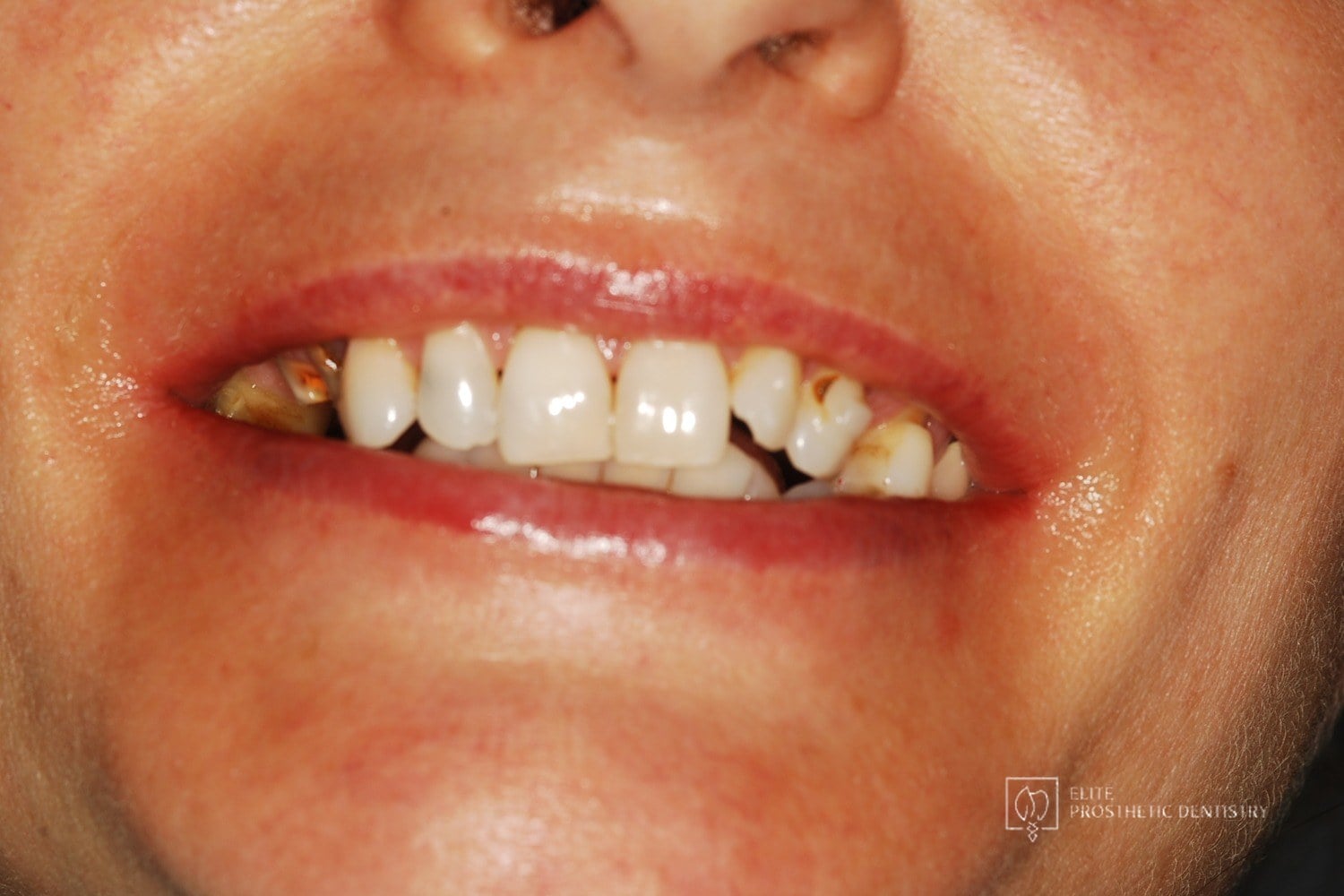 Before
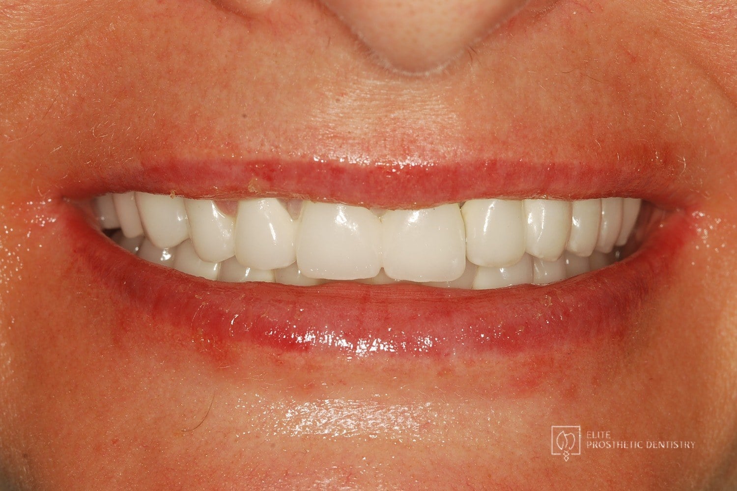 After
How Aging, Failing Dentistry Was Replaced with a Coordinated Full-Mouth Implant and Crown Reconstruction
The patient presented with throbbing upper-molar pain, long-standing infections, multiple root canals and unfinished prior treatment, generalized sensitivity, and several hopeless teeth.
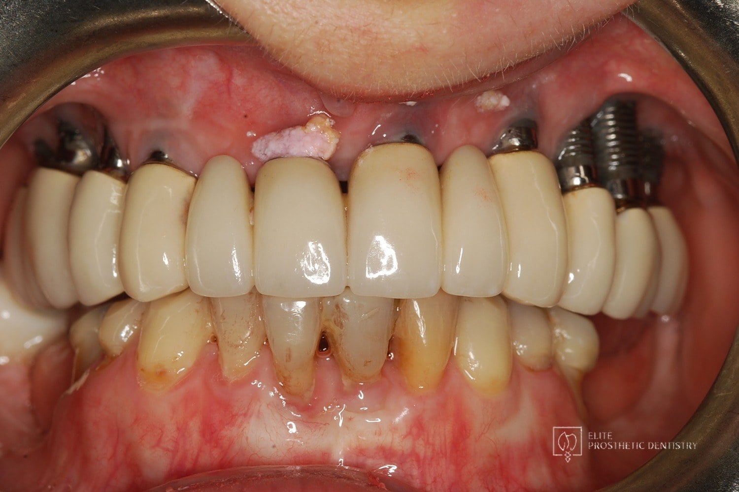 Before
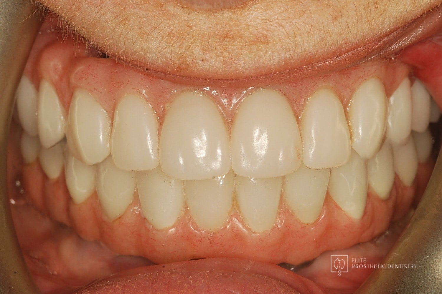 After
How Failing Implants and Severe Bone Loss Were Rebuilt Into a Stable Full-Arch Implant Reconstruction
The patient presented with significant complications from previously placed implants, severe bone loss, failing posterior implants, and an unesthetic full-arch bridge. Both arches required full reconstruction, with the upper arch showing the most advanced bone loss and instability.
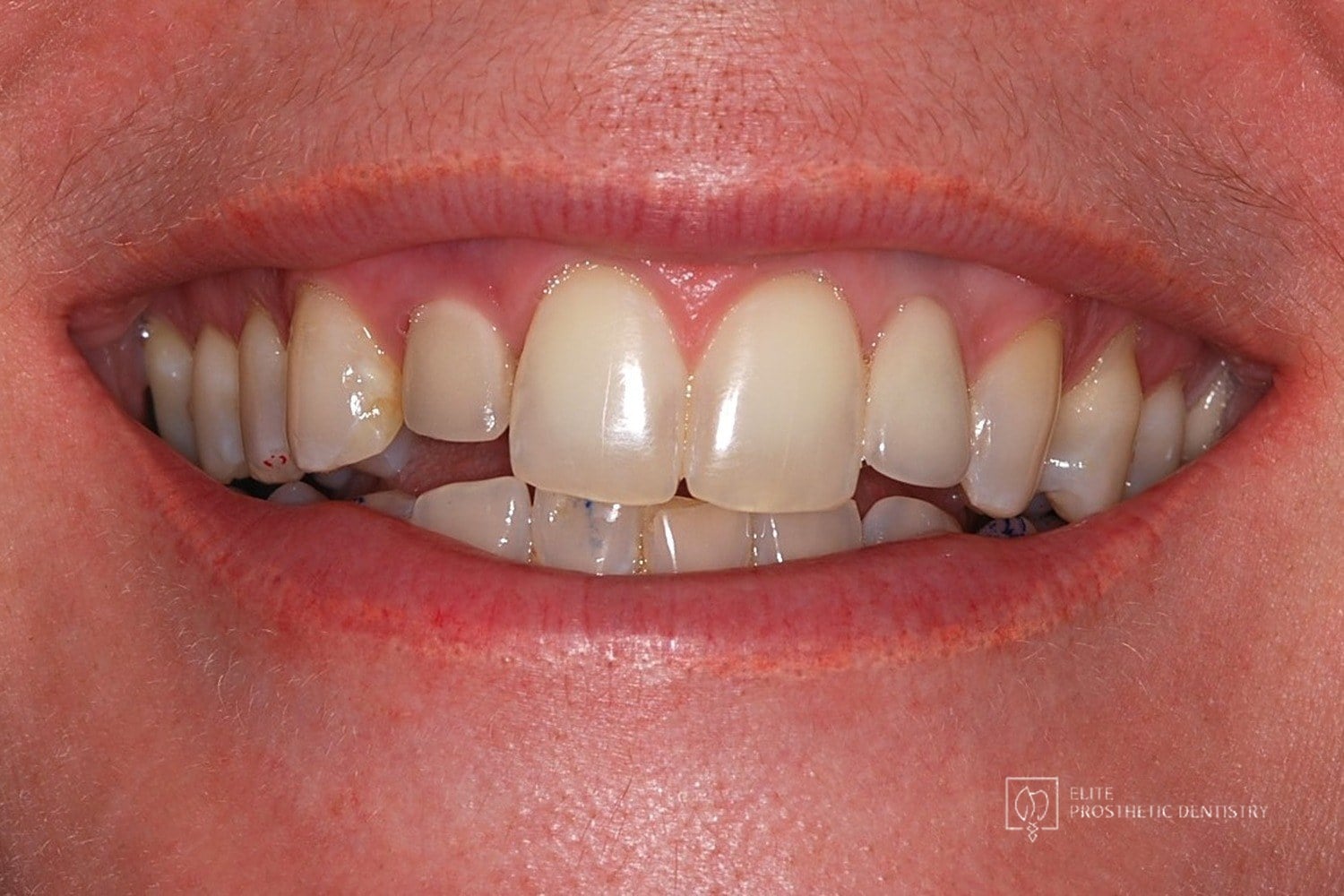 Before
 After
How Failing Lateral Incisor Implants Were Replaced with Custom Zirconia Bridges After Severe Bone Loss Made Replacement Implants Impossible
Comprehensive examination including CBCT imaging revealed significant bone loss surrounding implants that were placed outside the buccal plate, most likely due to placement without a CBCT-generated surgical guide. The implants were determined non-salvageable, and the alveolar ridge was too narrow to support replacement implants without extensive reconstruction.