
Front Tooth Broken at the Gum Line After a Bike Accident: Bone Graft, Guided Implant, and a Custom Crown
Intraoral view
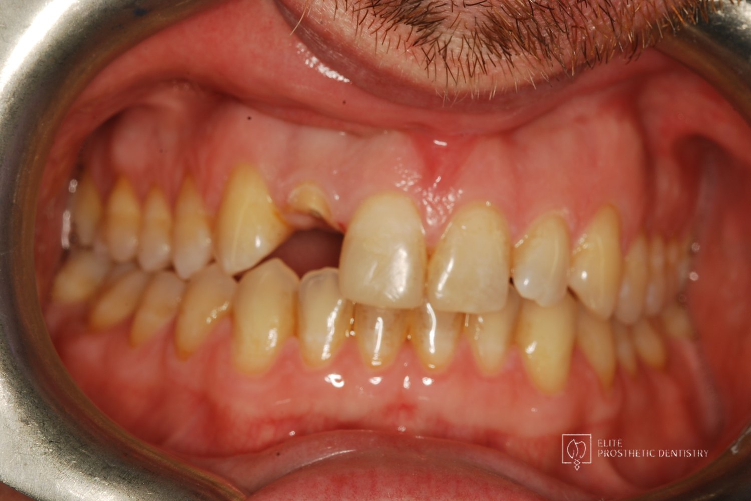 Before
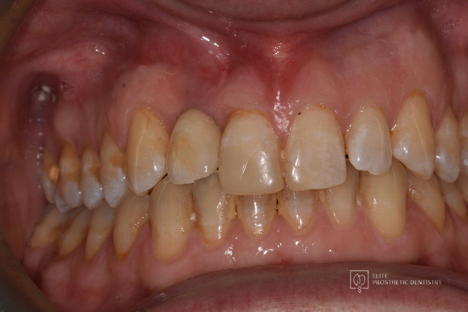 After
Close-up of the injured site
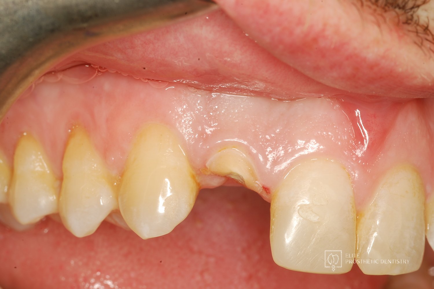 Before
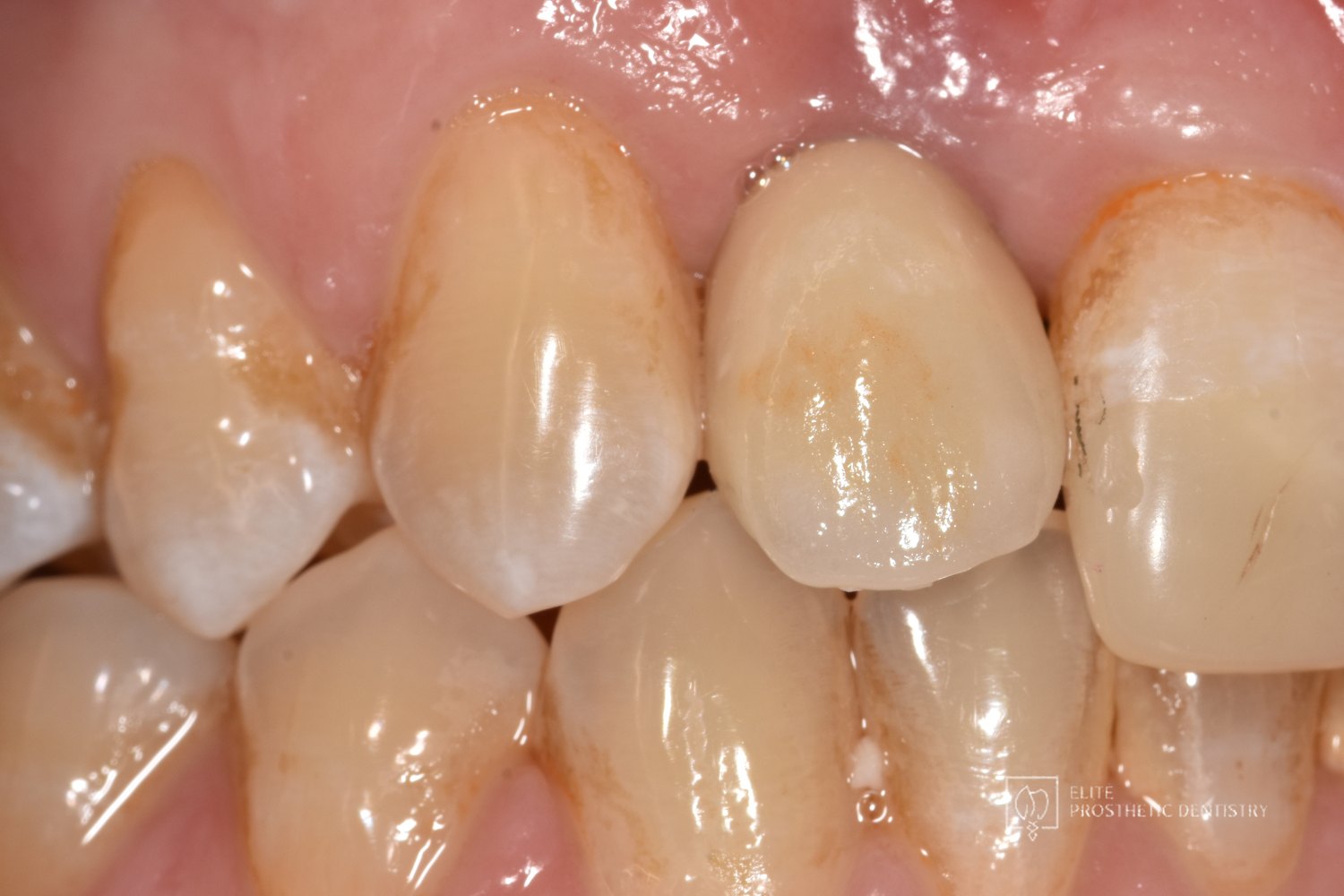 After
Smile before treatment
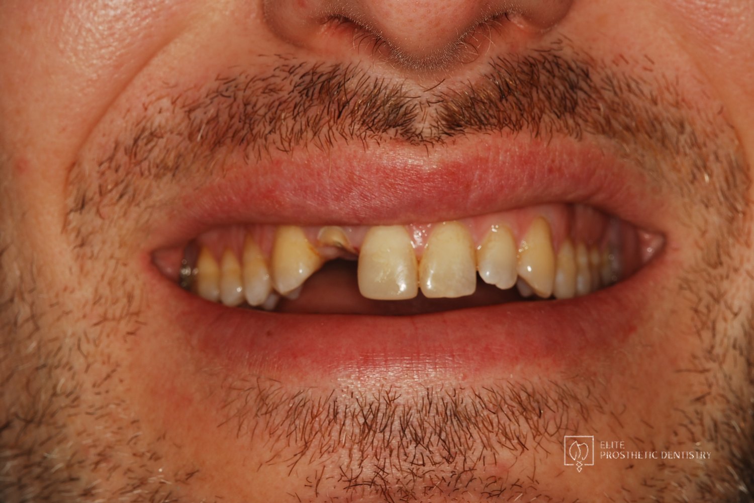
A front tooth lost to injury, rebuilt from the bone up
Replacing an upper front tooth broken at the gum line in Washington, DC for a patient who had injured it in a bike accident while traveling in Europe several years earlier. This documented case at Elite Prosthetic Dentistry used CBCT imaging to plan the extraction, bone grafting, and repair of a thin facial bone plate before a dental implant was placed and restored with a custom crown. Treatment was planned and completed by Gerald Marlin, D.M.D., M.S.D., a prosthodontist who plans implant surgery and the final restoration together.
Case at a Glance
- Treatment
- Single-tooth implant replacement of an upper front tooth after trauma, with bone grafting and a custom crown
- Approach
- CBCT imaging and full diagnostics, extraction with bone graft and repair of the facial bone plate, staged healing, implant placement, custom crown
- Case Type
- Single-tooth implant / Esthetic-zone replacement after trauma
- Patient Concern
- An upper front tooth injured in a bike accident abroad and patched temporarily for years had broken at the gum line; he was wearing a temporary partial and wanted a permanent solution.
- Decision Point
- Place an implant into a site with a thin, damaged facial bone plate, or rebuild the bone and gum shape first so the implant and crown could look and last like a natural tooth.
- Outcome
- A front tooth that looks like it belongs, supported by rebuilt bone and natural gum contour rather than compromised by the injury.
- Why It Matters
- Shows why a front-tooth implant after trauma begins with the bone and gum architecture, because the final crown can only look as good as the foundation under it.
The presenting condition
A few years before he came to the practice, this patient had a bike accident while traveling in Europe. The front tooth took the impact and was managed at the time with temporary repairs, which held for a while. Then the tooth broke at the gum line. He arrived wearing a temporary partial to fill the gap and hoping an implant could give him a permanent answer. The close-up photographs on this page show what the injury had left: very little tooth above the gum, and a facial bone plate that imaging showed to be shallow over the root.
Clinical Findings
- →Upper front tooth broken at the gum line following an earlier bike-accident injury
- →Years of temporary repairs that had reached the end of what they could do
- →A shallow facial bone plate over the site, documented on CBCT imaging
- →A visible gap in the smile line, managed with a temporary partial
- →A patient who wanted a permanent, natural-looking replacement rather than another interim fix
Why the bone and gum shape came before the implant
A front tooth sits in the thinnest bone in the mouth, and trauma makes that bone thinner still. If an implant goes into a site with a shallow or damaged facial plate, the bone can recede over time, the gum follows it, and the crown ends up looking long, dark at the neck, or simply different from its neighbor. For a tooth this visible, that is not an acceptable risk. The predictable path is to remove the damaged root carefully, graft the socket, rebuild the facial plate so the ridge regains its natural shape, allow it to heal, and then place the implant in bone that will support both the crown and the gum contour around it for the long term. The surgical sequence, the bone foundation, and the final crown are planned together before anything is done, which is what makes the result look like a tooth rather than a repair.
The treatment plan
The plan was built from CBCT imaging and full diagnostics so that each phase served the final crown.
-
1
CBCT imaging and diagnostics
A cone beam CT scan and complete records documented the remaining root, the thickness of the facial bone plate, and the position the implant would need to occupy for a natural-looking crown.
-
2
Extraction and bone grafting
The damaged tooth was removed with care to preserve the surrounding bone, and the socket was grafted to maintain the volume of the ridge. See bone grafting for how this protects the site.
-
3
Repair of the facial bone plate
The shallow facial plate was rebuilt so the gum would have natural support and the implant would have solid bone on its most visible side.
-
4
Implant placement after healing
Once the grafted site had matured, a dental implant was placed in the position planned from the scan. See precision implant placement.
-
5
Custom crown
The implant was restored with a custom crown designed to match the shape, color, and contour of the neighboring front teeth. The restoration was fabricated in direct coordination between Dr. Marlin and the practice's in-house laboratory to control fit, contour, and esthetics.
The outcome
The completed result replaced a tooth that had been patched for years with an implant-supported crown standing in rebuilt bone and natural gum contour. The retracted and close-up views at the top of this page show the change at the site itself: no gap, no temporary partial, and a crown that reads as one of the patient’s own teeth. The goal was never only to fill the space; it was to rebuild the support, shape, and strength the smile needed so the result would hold.
Result Highlights
- ✓CBCT imaging documented the shallow facial bone plate before any surgery
- ✓Extraction and grafting preserved the ridge, and the facial plate was rebuilt for natural gum support
- ✓Implant placed after healing in the position planned for the final crown
- ✓Custom crown matched to the neighboring front teeth
- ✓A permanent replacement after years of temporary repairs
Who this case may sound familiar to
This story tends to resonate with patients in a few recognizable situations:
- You broke or lost a front tooth in an accident and have been living with temporary fixes.
- You want an implant for a front tooth and want it to look like a natural tooth, not a repair.
- You have been told the bone over a front tooth is thin and you want to understand what rebuilding it involves.
- You want the surgery and the final crown planned by the same specialist so nothing gets lost between offices.
- You want it done correctly once so the result is still right years from now.
If any of those describe where you are, a consultation with Dr. Marlin can give you the diagnostic picture and the specific plan for your case.
Frequently asked questions
Can a front tooth broken at the gum line be replaced with an implant?
Usually, yes, but the sequence matters. The remaining root has to be removed without damaging the thin facial bone, the socket is grafted, and any deficiency in the facial plate is rebuilt before the implant is placed. Skipping those steps risks bone and gum recession that shows on a front tooth.
Why is CBCT imaging important for a front-tooth implant?
A cone beam CT scan shows the thickness of the facial bone in three dimensions, which a standard x-ray cannot. For a front tooth, that measurement decides whether grafting is needed, how the site should be rebuilt, and exactly where the implant should go so the crown looks natural.
How long after an injury can a front tooth still be replaced with an implant?
There is no fixed deadline. Years of temporary repairs or a missing tooth change the bone and gum, but those changes can usually be rebuilt with grafting. The evaluation looks at what bone remains and plans the reconstruction from there.
What does a prosthodontist do differently with a single front-tooth implant?
A prosthodontist plans the final crown first: its shape, its gum line, and how it will sit against the neighboring teeth. The implant position, the grafting, and the healing timeline are then chosen to serve that crown, rather than the crown being made to fit wherever an implant happened to be placed.
More about the work behind this case
This case combines a dental implant with bone grafting and guided placement in the esthetic zone. The staged approach reflects the practice’s philosophy on planning the endpoint first, and the crown was made with the in-house laboratory.
Elite Prosthetic Dentistry treats patients from across the DMV including Bethesda, Chevy Chase, McLean, Arlington, Potomac, and Great Falls, and from much further out for cases that warrant the trip.
Related Services
Related Patient Success Stories
Explore similar patient success stories demonstrating our expertise in advanced prosthetic dentistry.
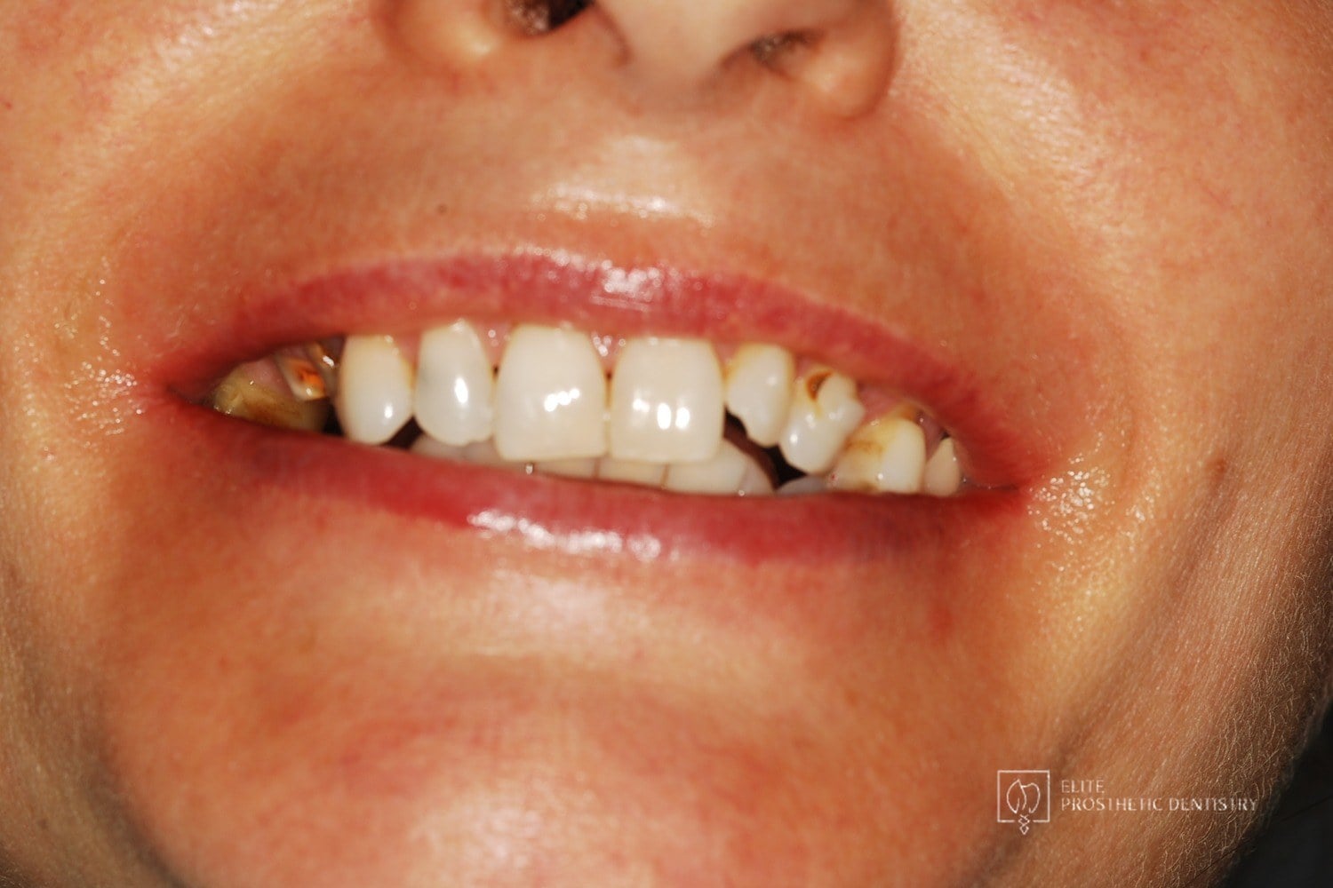 Before
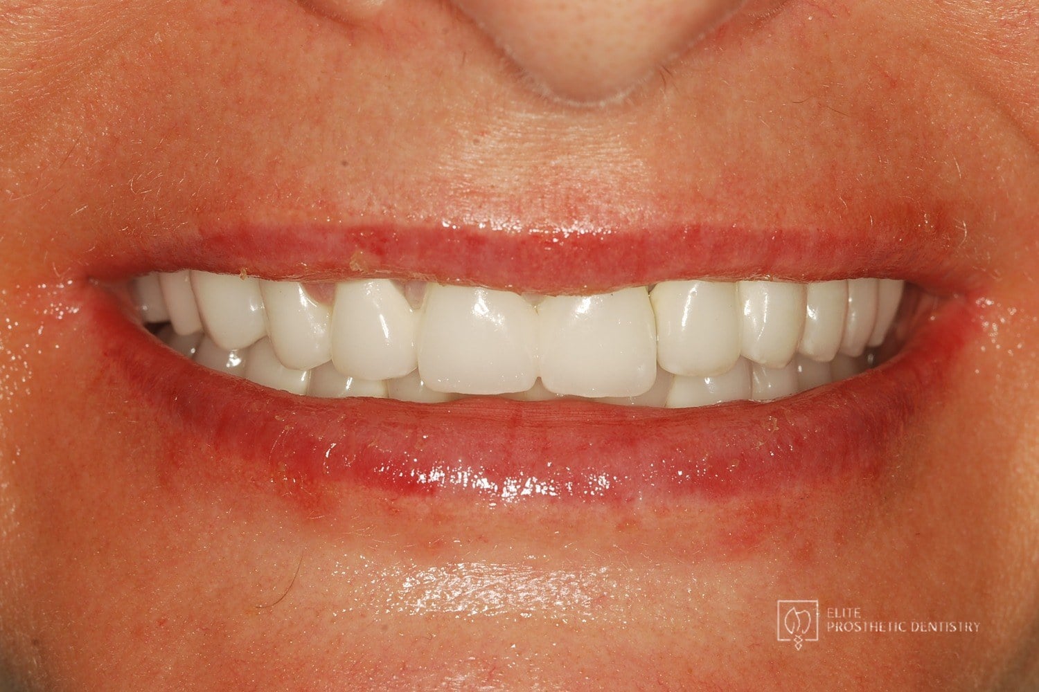 After
How Aging, Failing Dentistry Was Replaced with a Coordinated Full-Mouth Implant and Crown Reconstruction
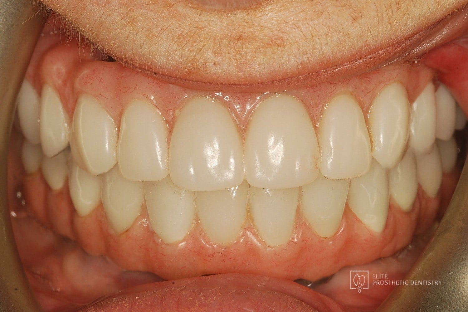
The patient presented with throbbing upper-molar pain, long-standing infections, multiple root canals and unfinished prior treatment, generalized sensitivity, and several hopeless teeth.
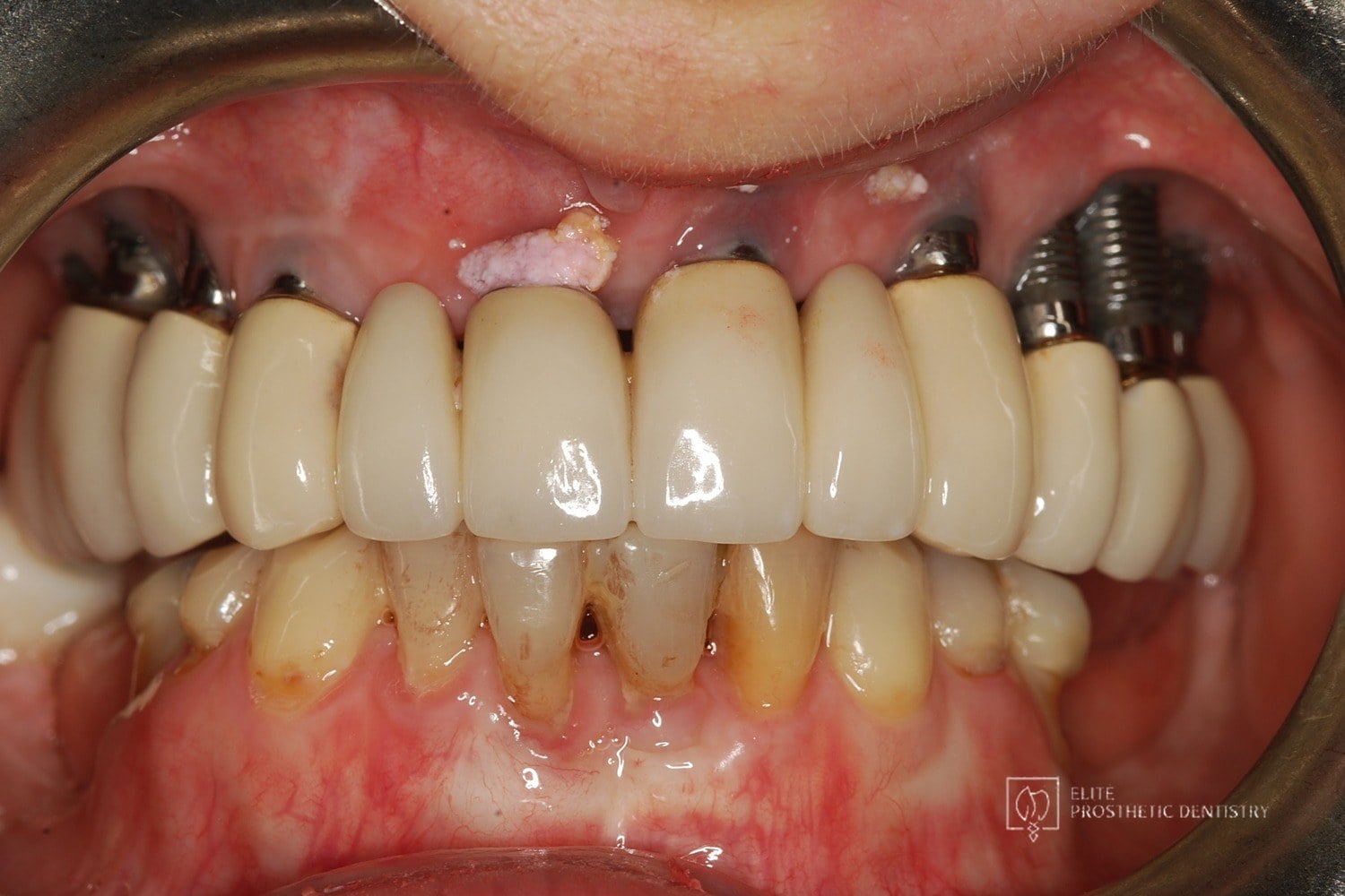 Before
 After
How Failing Implants and Severe Bone Loss Were Rebuilt Into a Stable Full-Arch Implant Reconstruction
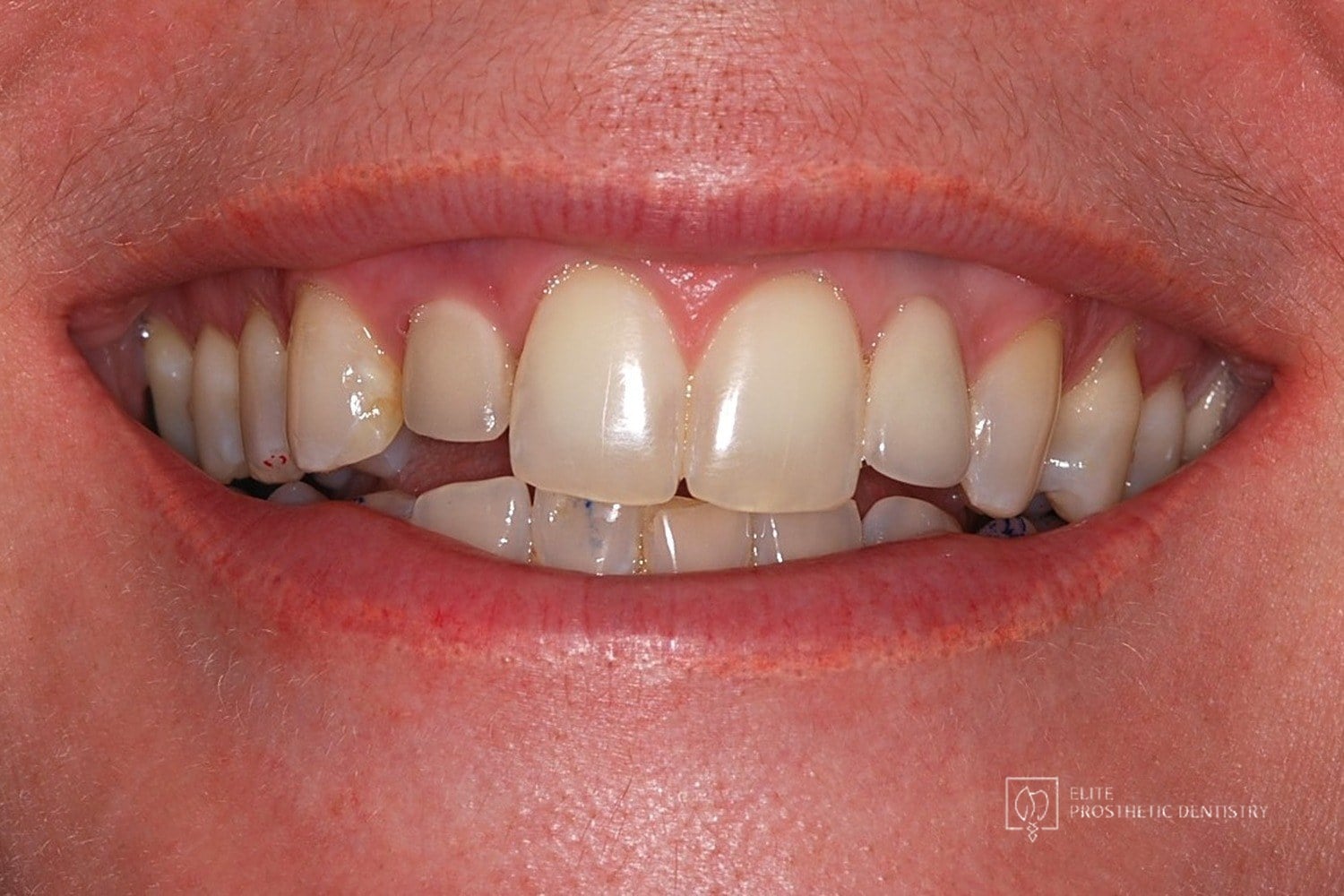
The patient presented with significant complications from previously placed implants, severe bone loss, failing posterior implants, and an unesthetic full-arch bridge. Both arches required full reconstruction, with the upper arch showing the most advanced bone loss and instability.
 Before
 After
How Failing Lateral Incisor Implants Were Replaced with Custom Zirconia Bridges After Severe Bone Loss Made Replacement Implants Impossible
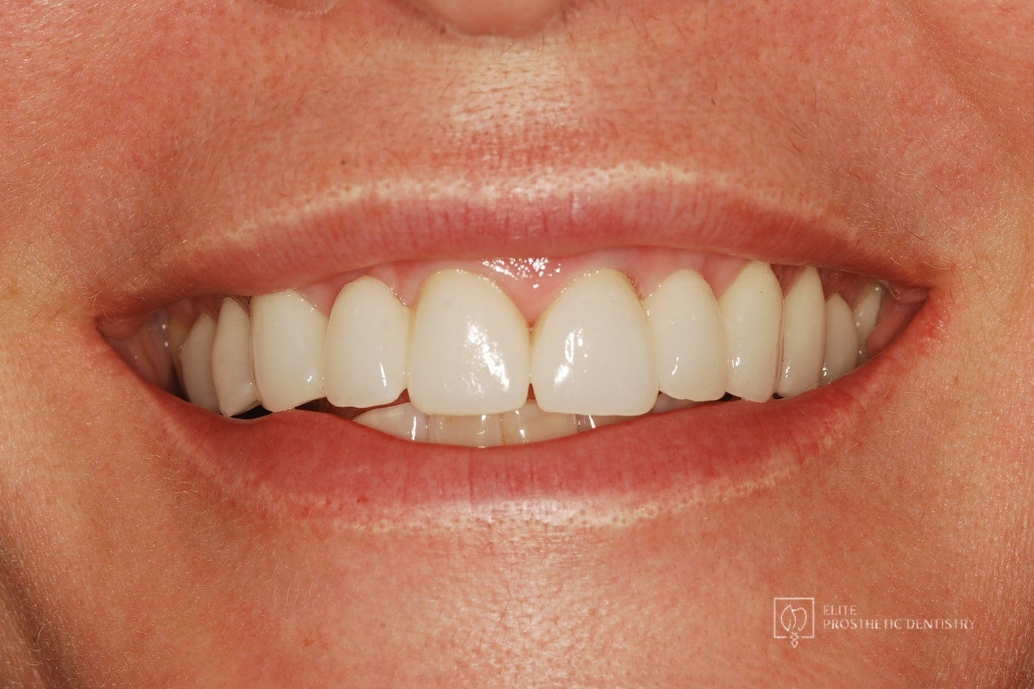
Comprehensive examination including CBCT imaging revealed significant bone loss surrounding implants that were placed outside the buccal plate, most likely due to placement without a CBCT-generated surgical guide. The implants were determined non-salvageable, and the alveolar ridge was too narrow to support replacement implants without extensive reconstruction.