Bone Grafting for Dental Implants in Washington, DC
Bone grafting at Elite Prosthetic Dentistry in Washington, DC. Dr. Marlin restores jawbone structure to support dental implants.
Restoring Bone Volume for Stronger, Longer-Lasting Implant Results
Why Is Bone Grafting Needed for Implants?
Bone grafting plays a critical role in implant success by rebuilding areas of the jaw that have lost volume or density. When teeth are missing, the jawbone in that area can begin to shrink, compromising the foundation needed for implant stability. Grafting allows us to reinforce the bone with natural or synthetic material so it can safely support a dental implant.
How Bone Grafting Works
During the grafting procedure, Dr. Marlin adds carefully selected grafting material to areas of bone loss in your jaw. This material may come from your own body (autograft), a donor (allograft), animal sources (xenograft), or be entirely synthetic (alloplast). Over time, your body naturally integrates this graft with your existing bone, creating a solid, healthy base for future implant placement.
Types of Bone Grafting Procedures We Offer
We perform several forms of grafting, depending on the location and severity of bone loss:
- Socket Preservation: Placed immediately after a tooth extraction to prevent bone shrinkage.
- Ridge Augmentation: Rebuilds the height or width of the jawbone for future implants.
- Sinus Lift: Adds bone to the upper jaw near the sinuses when there isn’t enough vertical height for implants.
- Block Grafting: Uses a small section of bone to rebuild larger areas of bone loss.
The Bone Loss Problem
When a tooth is lost, the bone that supported the tooth root begins to resorb, or shrink. This occurs because bone is living tissue that requires stimulation from tooth roots to maintain its volume. Without that stimulation, the body reabsorbs the bone as no longer necessary. This bone loss can be substantial, especially if a tooth has been missing for several years.
Bone loss can make implant placement impossible without grafting. It can also compromise the esthetic outcome if significant bone is missing in the front of the mouth. The gum line may appear lower, and teeth may appear longer than natural. It can affect your ability to wear a well-fitting denture. And severe bone loss can change your facial appearance, making you appear older.
Understanding Osseointegration
The process by which bone integrates with an implant is called osseointegration. In this biological process, bone cells grow directly onto the titanium implant surface, creating a strong, direct bond. This integration is what gives dental implants their stability and durability.
For osseointegration to occur successfully, the bone must be adequate in both quantity and quality. Bone quantity is the amount of bone present, measured in width and height. Bone quality refers to the density and structure of the bone. Dense bone with good blood supply typically integrates with implants more reliably and quickly. Less dense bone may require longer healing periods.
Bone Grafting Materials
Dr. Marlin may recommend different grafting materials depending on your specific situation.
Autograft: This is bone harvested from your own body, typically from the jaw or chin area. Autografts are considered the gold standard because they contain living cells that actively participate in bone formation. Your own bone tissue is the least likely to be rejected and heals the most quickly. However, harvesting requires an additional surgical site.
Allograft: This is bone harvested from a donor, usually from a bone bank. The donor bone is processed and sterilized. It provides structure for new bone formation but does not contain living cells. Allograft is effective, eliminates the need for a second surgical site on your body, and is widely used in implant dentistry.
Xenograft: This is bone harvested from an animal source, typically bovine (cow) bone. The bone is processed to remove all cellular material, leaving only the mineral structure. Xenograft provides excellent structural support and is particularly useful for ridge augmentation procedures.
Alloplast: These are entirely synthetic bone substitutes, created from biocompatible materials like calcium phosphate or hydroxyapatite. Alloplasts are convenient and effective for many grafting situations. Some are absorbable, while others are designed to remain as permanent scaffolding for new bone.
Dr. Marlin will discuss which material is most appropriate for your specific situation during your consultation.
Types of Bone Grafting Procedures We Offer
We perform several forms of grafting, depending on the location and severity of bone loss:
Socket Preservation: When a tooth is extracted, the empty socket can be filled with grafting material immediately. This preserves bone volume and reduces the amount of bone loss that would otherwise occur over the following months. This procedure is often performed at the time of extraction, preparing the site for future implant placement.
Ridge Augmentation: The alveolar ridge is the bone that supports your teeth. When multiple teeth are missing, the ridge can become narrow or low. Ridge augmentation rebuilds the height or width of the ridge to support implants and improve esthetics. This procedure may be performed before implant placement or in conjunction with it.
Sinus Lift: In the upper back jaw, the maxillary sinus is a hollow space above the tooth roots. When back upper teeth are missing, the sinus can expand downward, reducing the vertical height of bone available for implants. A sinus lift procedure elevates the sinus membrane and adds bone graft material underneath, creating space for implant placement.
Block Grafting: For large areas of bone loss, a block of bone may be harvested from an area like the back of the jaw or chin. This block is positioned and stabilized to rebuild the contours of the ridge. Block grafting is particularly useful for comprehensive full mouth reconstruction cases.
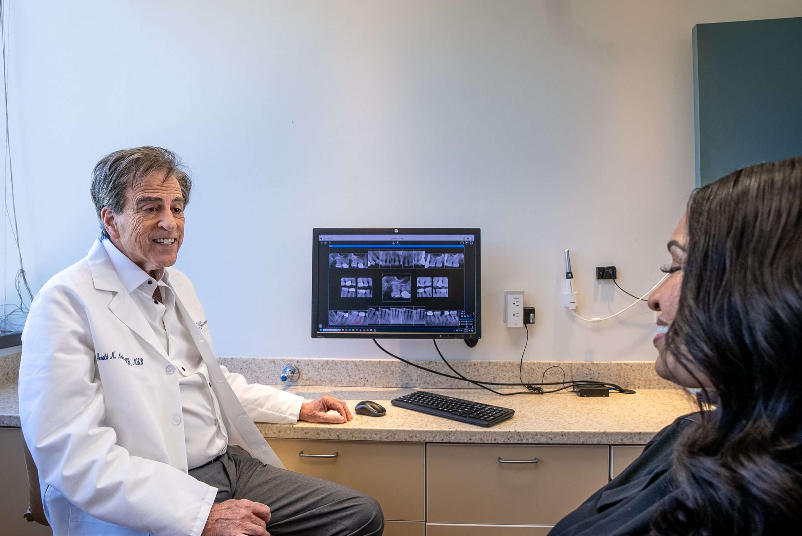
What to Expect From Treatment
Dr. Marlin and our team use advanced digital imaging and CBCT scans to precisely plan your bone graft. Most procedures are performed under local anesthesia with sedation if desired. After surgery, patients can expect mild swelling and discomfort for a few days. Healing typically takes a few months, but this added step leads to stronger, longer-lasting implant outcomes.
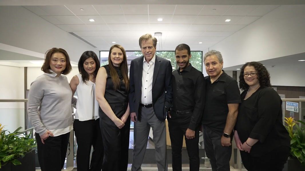
Why Choose Dr. Marlin for Bone Grafting
Bone grafting requires sophisticated surgical skills and a deep understanding of bone biology. Dr. Marlin has completed thousands of bone grafting procedures throughout his 40-year career. His experience allows him to select the most appropriate techniques and materials for your unique situation. His success rates with bone grafting are excellent, and many of his patients have proceeded successfully to implant placement after grafting.
The Long-Term Perspective
While bone grafting adds time and cost to your implant treatment, it often enables implant placement that would otherwise be impossible. The stronger foundation created by grafting typically results in higher implant success rates and longer implant longevity. For patients pursuing full mouth dental implants, full-arch options like All-on-4® or All-on-6®, or even a single implant crown, adequate bone is the foundation of a lasting result. Over the course of your lifetime, this translates to a superior outcome and better value than alternative tooth replacement options.
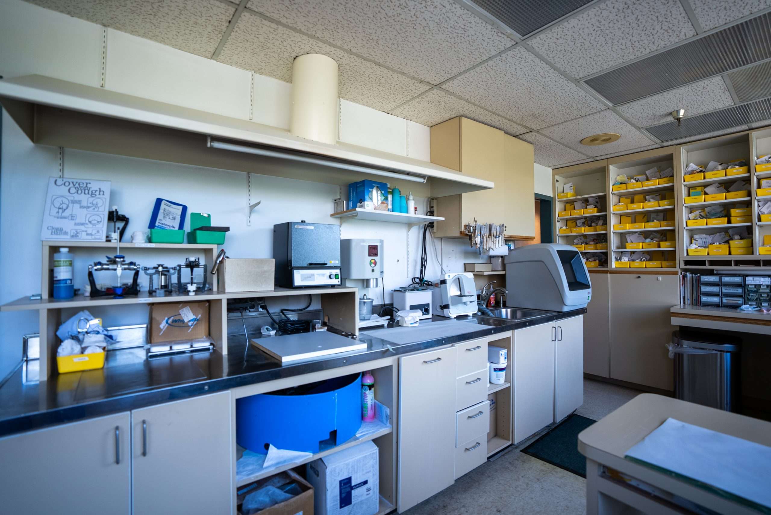
After Bone Grafting: Your Next Steps
Bone grafting creates the foundation for successful precision implant placement. Once healing is complete, Dr. Marlin can place dental implants that are restored with custom-crafted crowns fabricated in our in-house laboratory. Patients who have experienced implant failure often require bone grafting before replacement implants can be placed. Sedation dentistry is available to ensure comfort during grafting procedures.
Frequently Asked Questions
What does bone grafting do?
Bone grafting creates enough healthy bone volume to support a dental implant where bone loss has occurred.
Is bone grafting painful?
Most patients report only mild discomfort after the procedure, similar to a tooth extraction, and it’s managed with over-the-counter or prescribed pain relief.
How long does bone grafting take to heal?
Healing usually takes 3 to 6 months, depending on the type of graft and your individual healing response.
What are the risks of bone grafting?
Risks are minimal but can include swelling, infection, or graft rejection. Dr. Marlin’s precise technique helps reduce these risks significantly.
Can bone grafting help even with severe bone loss?
Yes. Bone grafting makes implants possible even in cases of severe bone loss, restoring the foundation needed for success.
Serving Patients From
Maryland
Bethesda (Bone Grafting) | Bethesda (Sinus Lift) | Cabin John | Chevy Chase | Kensington | North Bethesda | Potomac | Rockville | Silver Spring
Washington, DC
Cleveland Park | Dupont Circle | Foxhall | Georgetown | Kalorama | Palisades | Spring Valley | Tenleytown | Woodley Park
Virginia
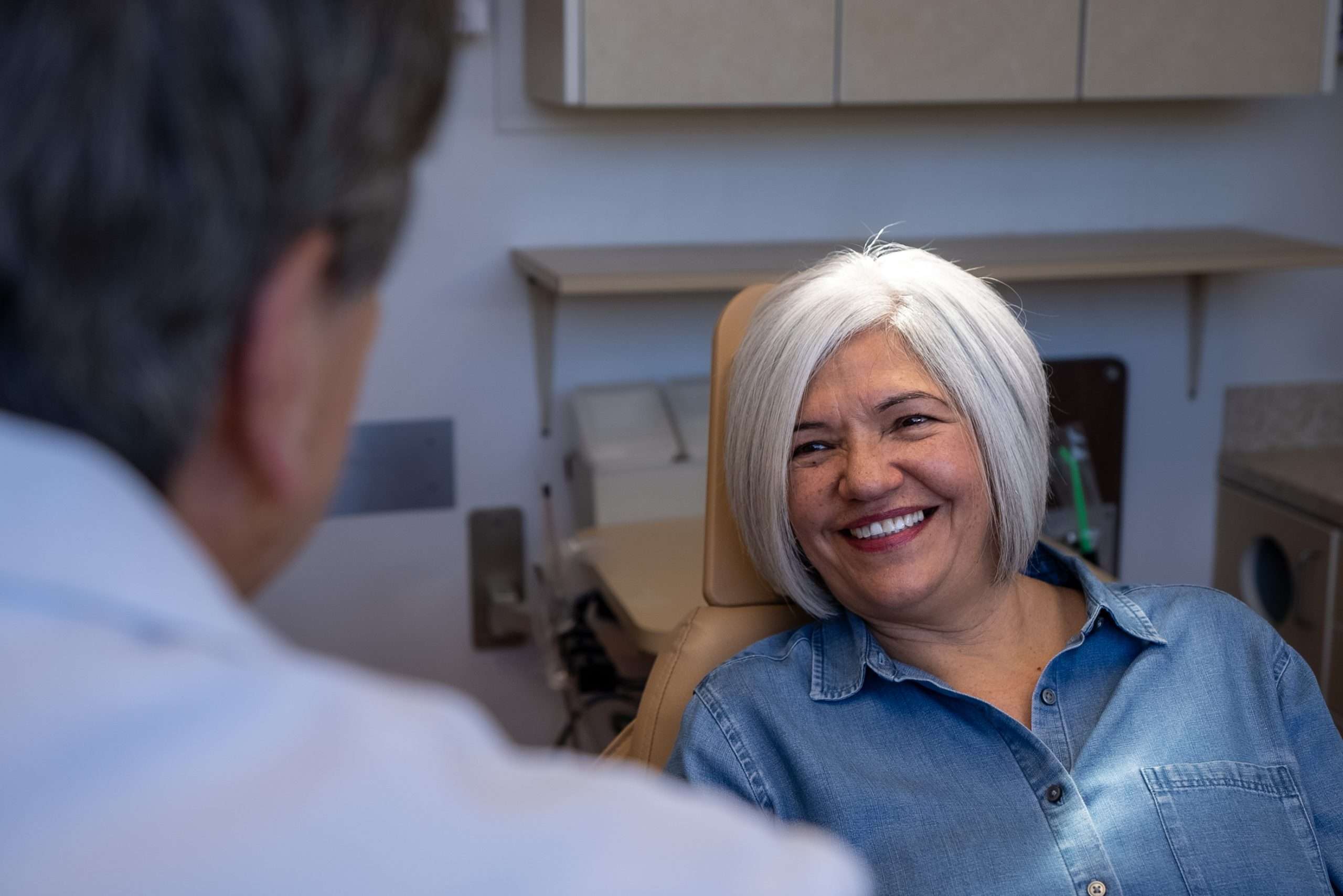
Your Best Smile Is Within Reach
Request a specialist consultation with Dr. Marlin to discuss your situation and the most appropriate path forward.
Related Patient Success Stories
Explore similar patient success stories demonstrating our expertise in advanced prosthetic dentistry.
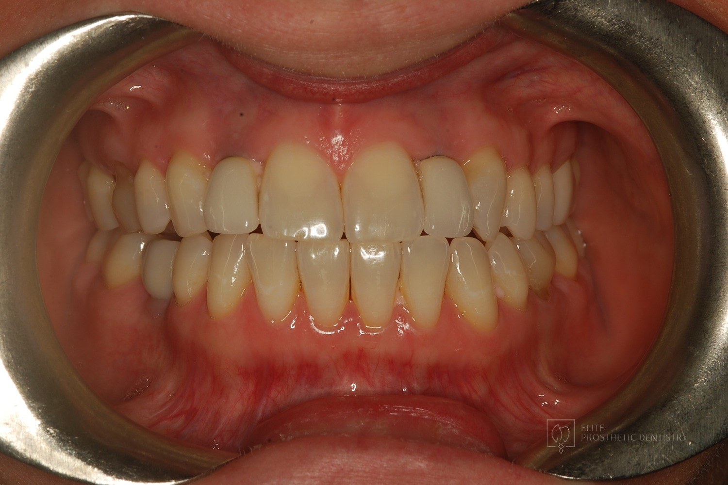 Before
 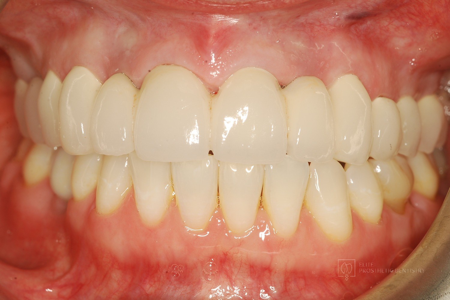 After
After How Older Implant Crowns Were Redesigned for a Better Bite and More Natural Appearance
The patient came in after years of living with implant-supported crowns placed more than twenty years earlier that no longer looked or functioned well. CBCT evaluation, reviewed with a radiologist colleague, showed the implants had been placed too far to the buccal in very thin bone and could not support a healthy long-term restoration.
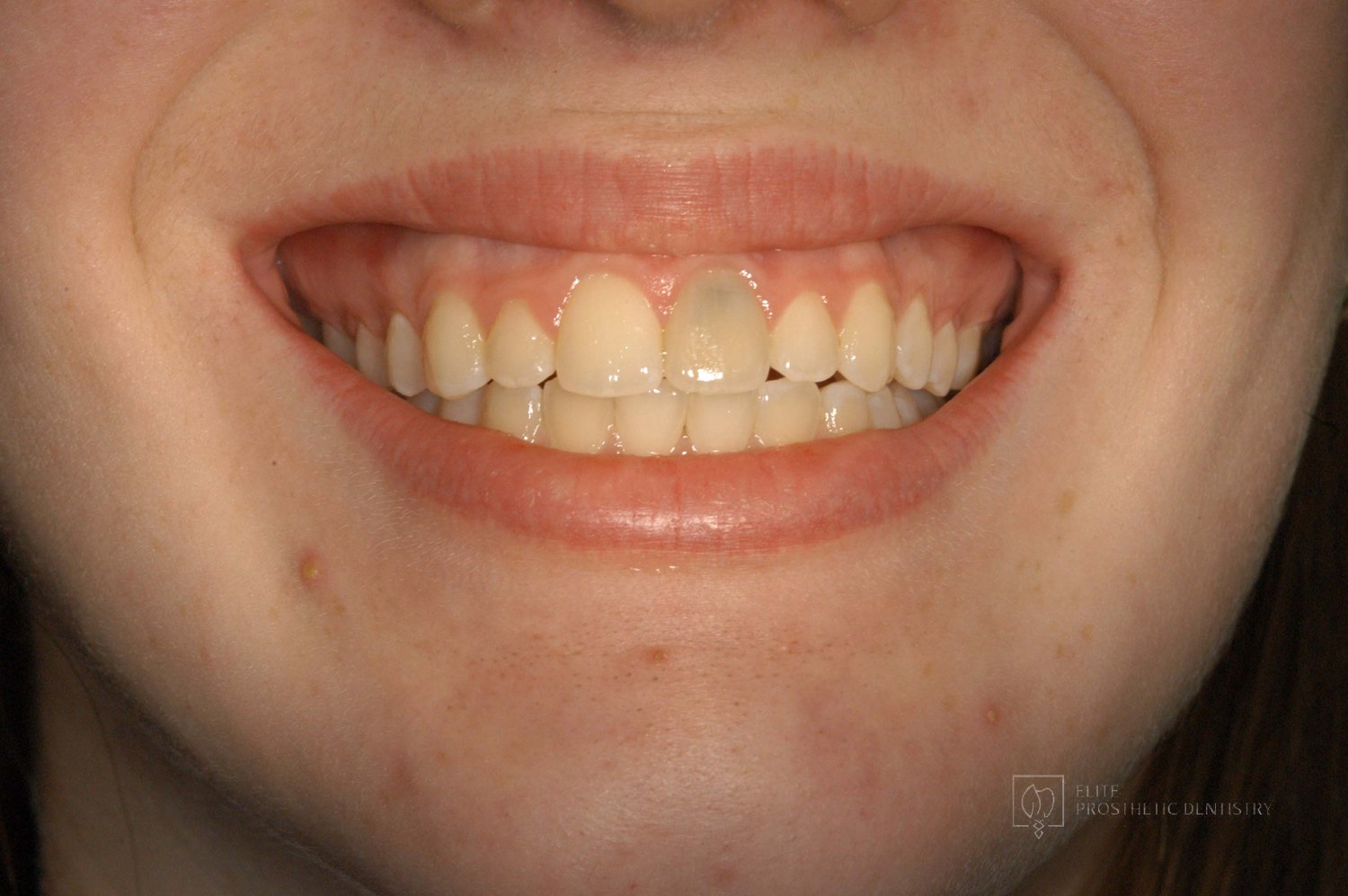 Before
 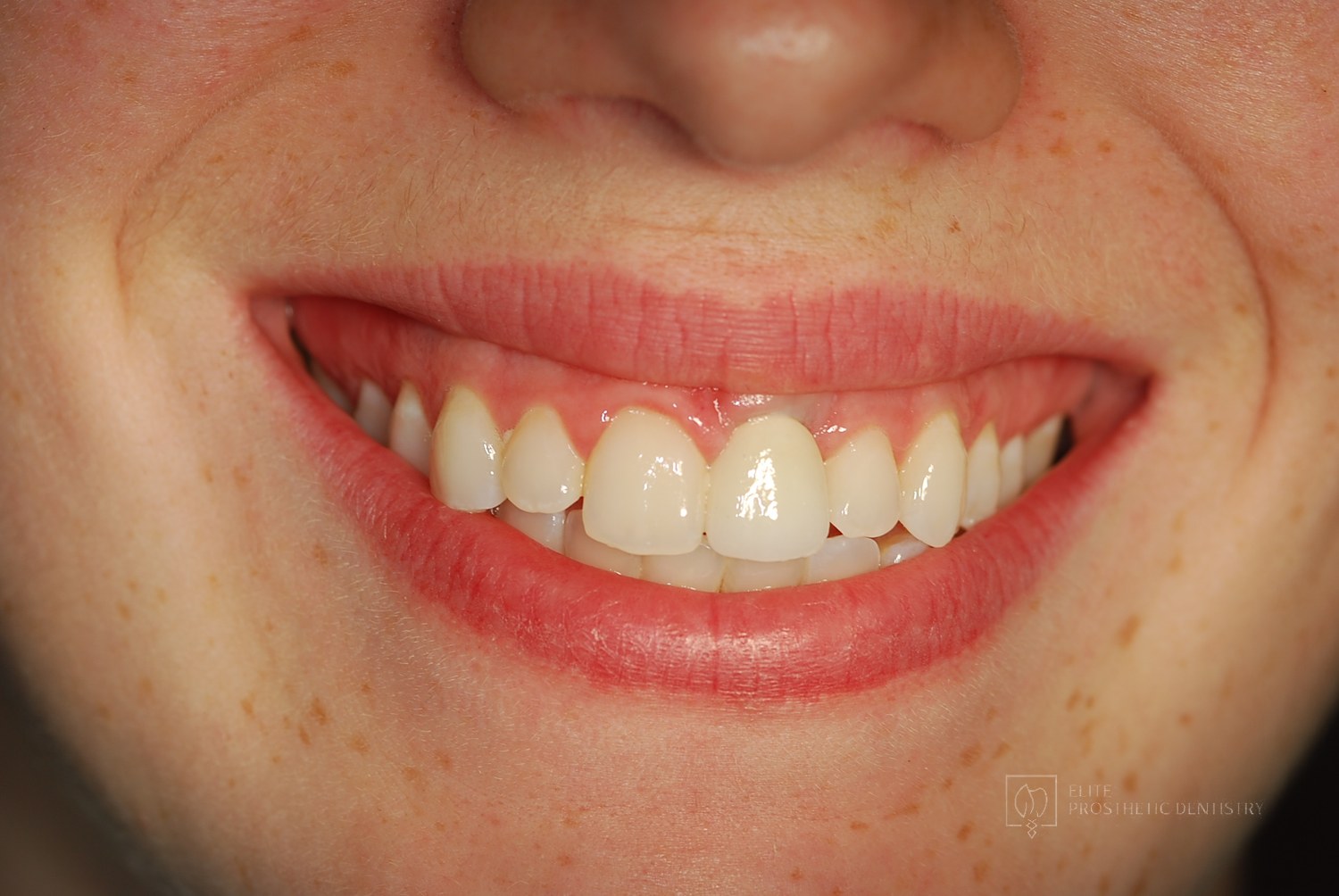 After
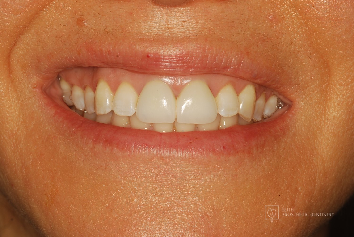
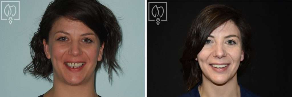
After How a Front Tooth Lost to Childhood Trauma Was Rebuilt with Bone Grafting and a Long-Lasting Implant
A teenager was referred by her father after earlier trauma left her upper left front tooth slowly failing from root resorption. She was still growing, so an immediate implant was the wrong move. The tooth had to be maintained to buy time, then replaced correctly once she reached skeletal maturity.
 Before
 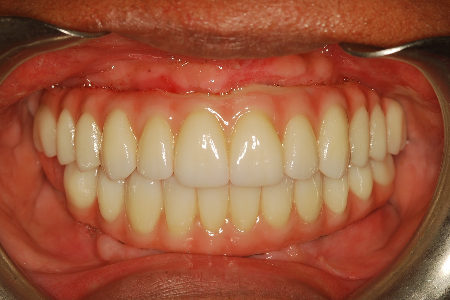 After
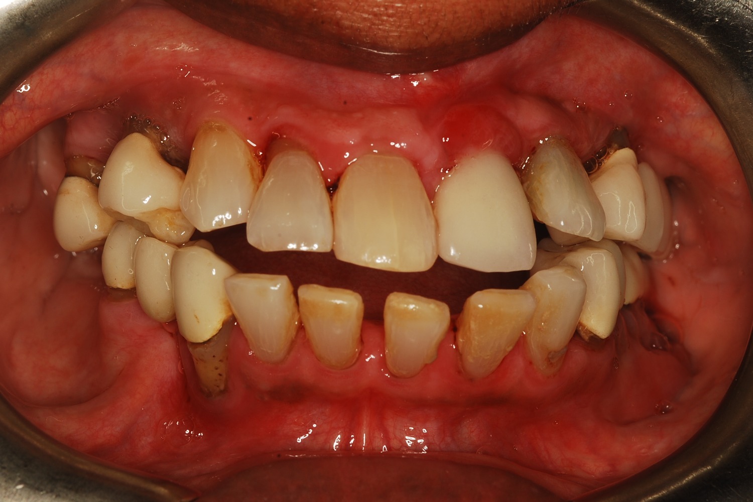
After How Severe Bone Loss and Bite Dysfunction Were Rebuilt with All-on-6 Implants and a Milled Zirconia Hybrid Prosthesis
The patient presented with severe bone loss, advanced periodontal disease, malocclusion, and a dysfunctional bite that required full-arch rebuilding.
 Before
 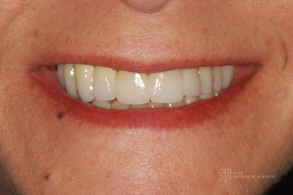 After
After Implant Supported Reconstruction: Failing Bridgework and Missing Back Teeth Rebuilt with Coordinated Specialist Care
Referred by another dental specialist with severe bone resorption on the upper left, multiple broken-down lower teeth requiring extraction, and failing lower back teeth that had left the bite without solid support. No single procedure, and no single provider working alone, could rebuild a situation this interconnected.
 Before
 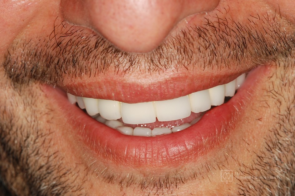 After
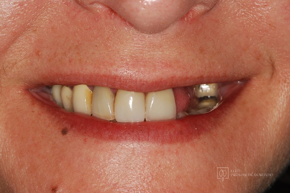
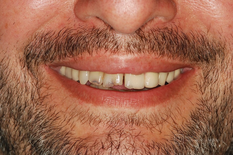
After Severe Restorative Breakdown Rebuilt with a Coordinated Full-Mouth Reconstruction
Multiple older restorations placed at different times over many years, broken-down teeth, a significant malocclusion, an asymmetrical smile, and two upper front teeth that could no longer be saved. No single repair could address a pattern this widespread.
 Before
 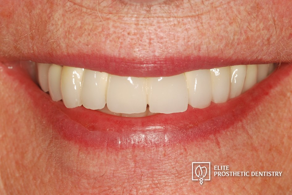 After
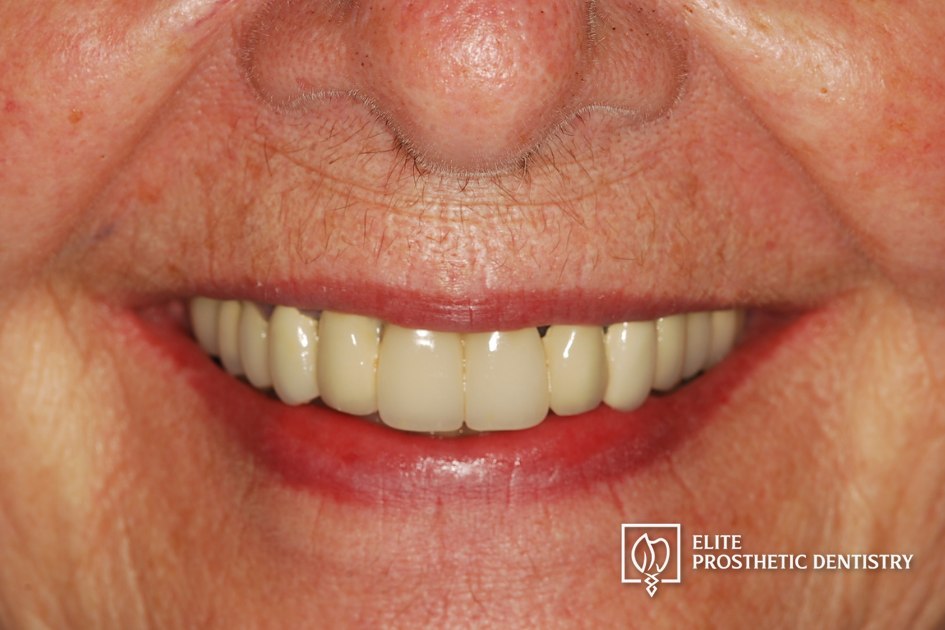
After How a Loose Upper Bridge and Aging Crowns Were Rebuilt with Staged Implant Reconstruction
A patient referred by her general dentist after years of aging dentistry no longer holding up. A loose upper bridge and crowns more than twenty years old, combined with the effects of advanced periodontal disease and severely compromised tooth abutments, required a staged surgical and restorative plan delivered with comfort planning at the same time.
Related Articles
Deepen your knowledge with additional insights on this topic.
Dental Implants If a Single Front Tooth Is Replaced with an Implant, Can It Look Natural?
Yes. See the four steps, with real case photos, that make a single front tooth implant indistinguishable from the natural tooth beside it. Washington, DC.
 Dental Implants
 What Is Precision Implant Placement (PIP)?
Precision Implant Placement plans each implant virtually on a CBCT scan, then delivers it with a custom surgical guide. See the three steps with real images.
 Dental Implants
 What Is the Ideal Surgical Guide for Precision Implant Placement?
Not all surgical guides are equal. The gold standard is CBCT-based: planned virtually in 3D, 3D printed, and seated on your teeth. A DC prosthodontist explains.
 Dental Implants
 When Should an Implant Not Be Done?
Healthy roots, thin bone, steep bony angles: a DC prosthodontist shows a real case where refusing implants was the right call, and what was done instead.
 Dental Implants
 What Is Staged Implant Therapy?
Staged implant therapy replaces failing teeth in phases while you keep fixed teeth the entire time. A DC prosthodontist walks through a real case in stages.
Dental Implants The Four Types of Dental Implants: Which One Is Right for You?
Endosteal, subperiosteal, zygomatic, and mini implants each solve a different problem. A DC prosthodontist explains the four types and who each one fits.
Bone Grafting for Dental Implants in Washington, DC Near You
Dr. Marlin provides bone grafting for dental implants in washington, dc services to patients throughout the Washington, DC metropolitan area. Select your community to learn more.
Ready to Transform Your Smile?
With 40+ years of experience and 3,900+ dental implants placed and restored, Dr. Marlin delivers results that last. Request a specialist consultation.