
The Single Anterior Crown
Documented before and after
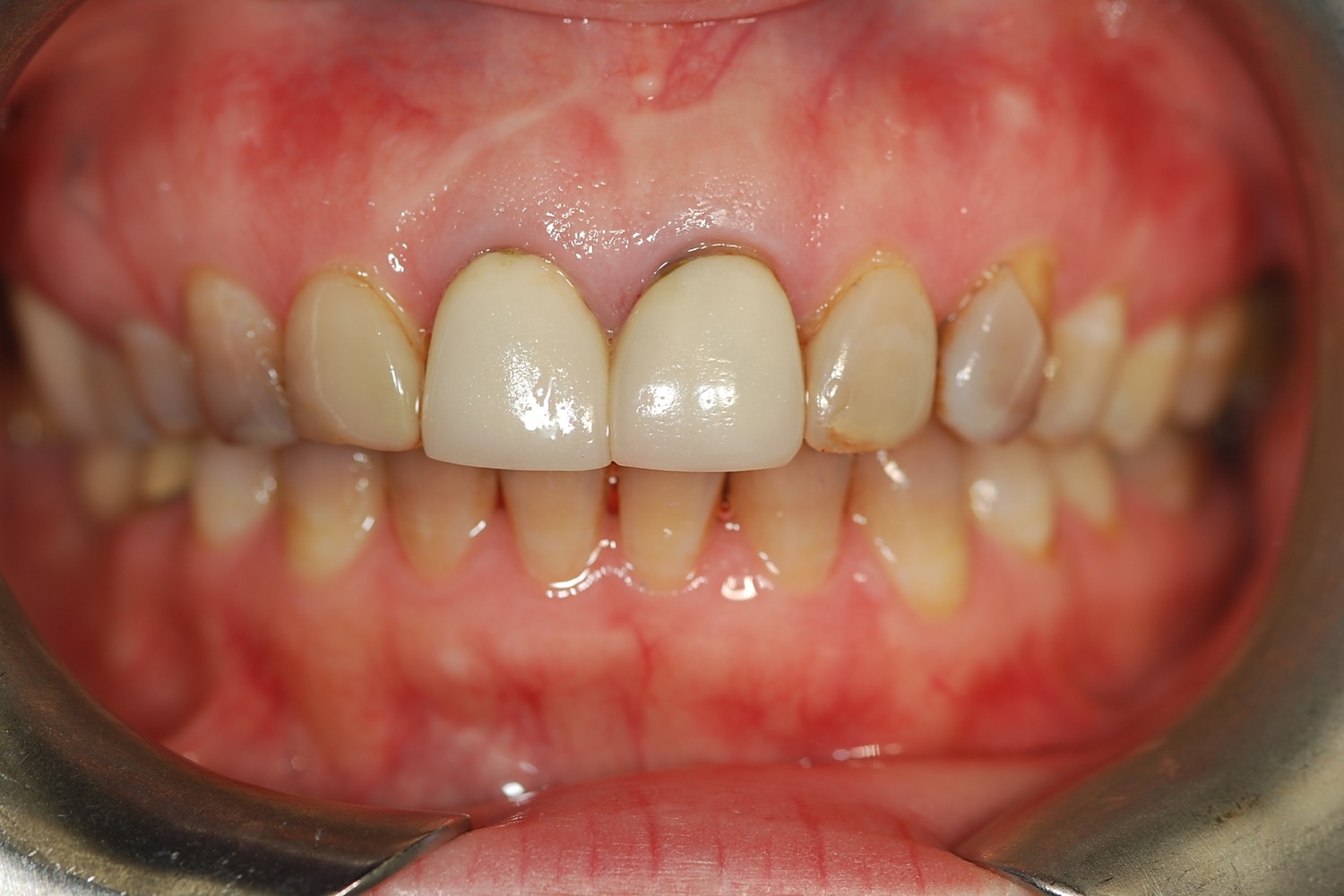
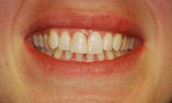 Before
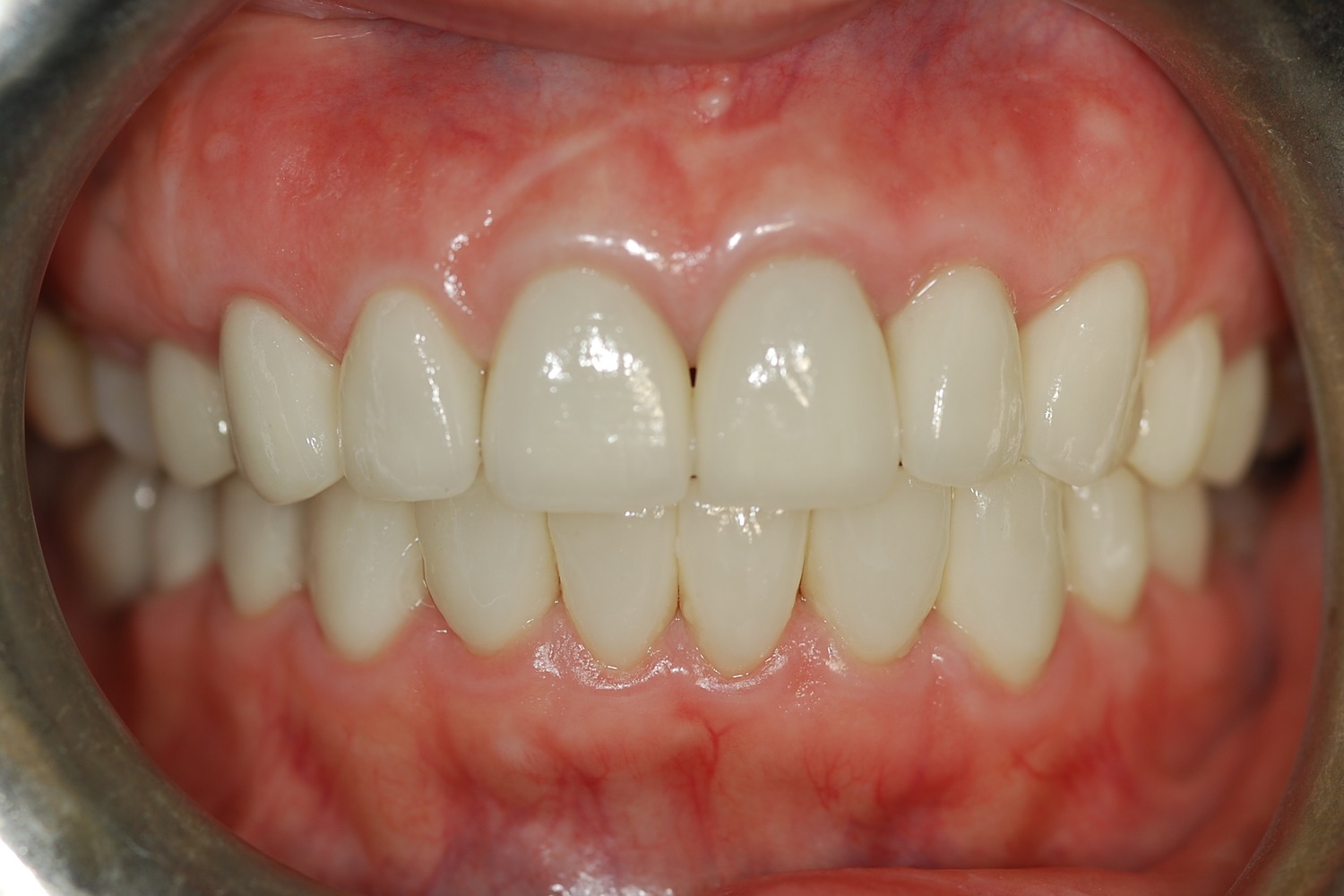
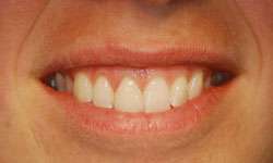 After
After
Additional documented view
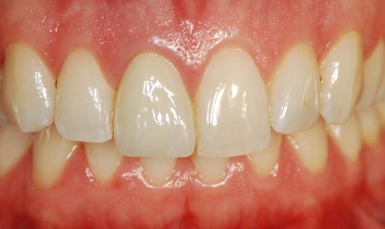
Final documented view
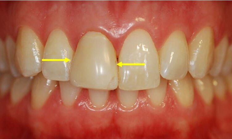
A single front tooth matched to its natural neighbor
Restoring a single front tooth in Washington, DC is one of the most esthetically demanding situations in restorative dentistry. This documented case at Elite Prosthetic Dentistry replaced a mismatched veneer on an upper central incisor with an all-ceramic crown that matches the natural tooth beside it, while resolving the gum inflammation around the old restoration. Treatment was planned and completed by Dr. Gerald Marlin, D.M.D., M.S.D., a prosthodontist focused on anterior esthetic restoration and complex restorative care.
Case at a Glance
- Treatment
- Single all-ceramic crown replacing a mismatched veneer on an upper central incisor
- Approach
- Whitening sequenced first, biocompatible contours for gum health, in-house laboratory fabrication
The presenting condition
The patient presented with a situation familiar in cosmetic dentistry: a veneer on her upper right central incisor whose shape, size, and proportions did not match her upper left central incisor. The veneer read as more square and artificial next to the natural, slightly tapered form of the adjacent tooth, and its shade was too opaque, without the translucency that characterized the natural incisor beside it. The mismatch was visible every time she smiled.
The concern was not only cosmetic. The veneer carried bulky contours, and its thickness and emergence angle were mechanically irritating the surrounding gum tissue, producing visible inflammation and swelling around the restoration. That tissue response looked unhealthy and created real discomfort for the patient.
Clinical Findings
- →Veneer on the upper right central incisor mismatched to the natural upper left central incisor
- →Shape more square than the slightly tapered form of the adjacent natural tooth
- →Bulky contours and an emergence angle irritating the gum tissue
- →Visible gingival inflammation and swelling around the restoration, with discomfort
- →Shade too opaque, lacking the translucency of the neighboring natural tooth
Why this case required prosthodontic-level planning
Replacing or restoring a single anterior tooth is one of the most esthetically challenging situations in restorative dentistry. With a natural tooth on one side and a restoration on the other, there is nowhere for the restoration to hide. It must match not just the color and basic shape of its neighbor, but the subtle contours, translucency characteristics, and light reflection properties that make a tooth look natural and alive. Small misses that would disappear in a full set of restorations are obvious when the comparison stands one tooth away.
This case added a biological dimension. Gum tissue responds to the contours of the restoration it surrounds, and tissue that is being mechanically irritated will stay inflamed no matter how diligent the patient’s hygiene is. The replacement had to be designed for the health of the gumline as much as for the appearance of the tooth, which meant the emergence profile below the gum mattered as much as the porcelain above it.
The decision behind the result: a crown instead of a new veneer, and whitening first
Two judgments shaped this outcome. The first was the choice of restoration. Dr. Marlin recommended replacing the veneer with an all-ceramic porcelain crown. A crown requires slightly more tooth preparation than a veneer, but it provides superior control over the final restoration’s characteristics, and in this case it allowed the contours to be redesigned so they would support healthy gum tissue instead of irritating it. The choice was decision-driven: not crown versus veneer in the abstract, but which restoration could deliver both symmetry and tissue health for this tooth.
The second judgment was about sequence. Before the new crown was fabricated, the patient’s other teeth were professionally brightened with teeth whitening treatment. Porcelain does not change color once made, so a crown built to match slightly yellowed enamel locks in that shade permanently. Whitening first established the final shade of the smile, and the crown was then fabricated to match the brightened teeth, preventing the mismatch that would otherwise have appeared later. It is a small step in the plan and a large one in the result.
The treatment plan
-
1
Evaluation of the mismatch and the tissue response
Assessment of the veneer's shape, shade, and contours against the natural central incisor, and of the inflammation in the surrounding gum tissue.
-
2
Professional whitening first
The surrounding natural teeth were professionally whitened before fabrication, so the new crown could be built to match the final, brightened shade of the smile.
-
3
Replacement with an all-ceramic crown
The veneer was replaced with an all-ceramic porcelain crown, chosen for the superior control it offered over shape, shade behavior, and tissue-friendly contours.
-
4
In-house laboratory design and fabrication
The practice's in-house laboratory matched the natural, slightly tapered shape of the adjacent incisor and engineered contours that emerge smoothly from beneath the gumline, with optimal translucency, internal color grading, and subtle surface texture.
-
5
Chairside integration of the final crown
The prosthodontic team worked directly with Dr. Marlin at the chair during delivery, confirming the integration of the new restoration with the tooth, the tissue, and the smile.
The outcome
The result exceeded the patient’s expectations. The new crown matches the shape, size, and color of the adjacent natural tooth, restoring complete visual symmetry to her smile. Its translucency and internal color grading make it very difficult to identify as a restoration at all.
Just as significant is what happened at the gumline. The crown’s biocompatible contours allowed the gingival tissue to become healthy and pink again, resolving the inflammation that had surrounded the previous restoration. The patient is pleased with the appearance, the comfort, and the health of her smile, and she can smile knowing the restoration looks natural and her gum tissue is well supported.
Result Highlights
- ✓New crown matches the shape, size, and color of the adjacent natural tooth
- ✓Complete visual symmetry restored between the two front teeth
- ✓Gum inflammation resolved, with healthy pink tissue around biocompatible contours
- ✓Whitening sequenced first so the crown was fabricated to the final shade
- ✓Translucency and internal color grading built in by the in-house laboratory
- ✓Final integration confirmed chairside by Dr. Marlin and the prosthodontic team
Who this case may sound familiar to
This story tends to resonate with patients in a few recognizable situations:
- You have one front-tooth crown or veneer that does not quite match its neighbor, and your eye goes straight to it in photographs.
- The gum around a front-tooth restoration stays red, puffy, or tender no matter how carefully you clean.
- You are considering whitening but worry an existing or planned restoration will no longer match afterward.
- You have been living with a front tooth that looks flat or opaque next to your natural teeth.
- You want a single, unforgiving restoration planned by a specialist and done correctly once.
If any of those describe where you are, a consultation with Dr. Marlin can establish the diagnostic picture and the specific options for your case.
Frequently asked questions
Why is a single front tooth the hardest restoration to match?
Because the natural tooth beside it sets the standard, and the comparison is permanent. The restoration must match color, shape, contour, translucency, and the way light reflects off the surface. In a full set of restorations, small deviations blend; next to a single natural incisor, they show.
How do you decide between a crown and a veneer on a front tooth?
Case by case. A veneer conserves more tooth structure, while a crown requires slightly more preparation but offers greater control over the final shape, shade behavior, and the contours that meet the gum tissue. The deciding factors include how much correction is needed, the condition of the tooth, and what the surrounding tissue requires to stay healthy.
Can a crown or veneer cause gum inflammation?
Yes. When a restoration’s contours are too bulky or its emergence angle presses into the surrounding tissue, the gum can become chronically inflamed and swollen even with good home care. Redesigning the restoration with biocompatible contours that support the tissue typically allows the gum to return to health.
Should I whiten my teeth before or after getting a crown?
Before. Porcelain does not change color once fabricated, so a crown made to match your current shade will no longer match if you whiten later. Whitening first establishes the final shade of the smile, and the crown is then built to match it.
Why see a prosthodontist for a single anterior crown?
Because every dimension of the restoration, from shade and translucency to contour and emergence profile, must be controlled at once, and the margin for error is close to zero. A prosthodontist is specialty-trained in exactly this kind of restorative design, and close collaboration with the laboratory is what turns that design into a tooth that reads as natural.
More about the work behind this case
This case brings together custom dental crowns, veneers judgment, and the practice’s in-house laboratory, all applied to a single unforgiving tooth. Designing for tissue health and long-term match, not just delivery-day appearance, is part of the practice philosophy behind anterior esthetic work here.
Elite Prosthetic Dentistry treats patients from across the DMV including Bethesda, Chevy Chase, McLean, Arlington, Potomac, and Great Falls, with a record of out-of-area patients traveling to the practice for complex restorative care.
Related Services
Related Patient Success Stories
Explore similar patient success stories demonstrating our expertise in advanced prosthetic dentistry.
 Before
  After
After How Aging, Failing Dentistry Was Replaced with a Coordinated Full-Mouth Implant and Crown Reconstruction
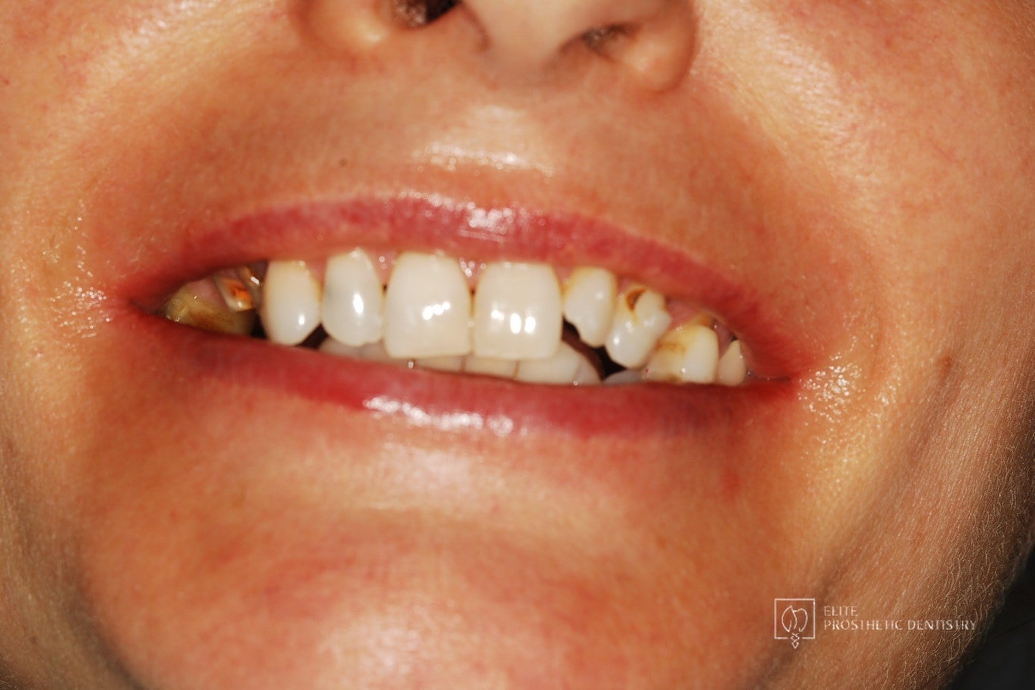
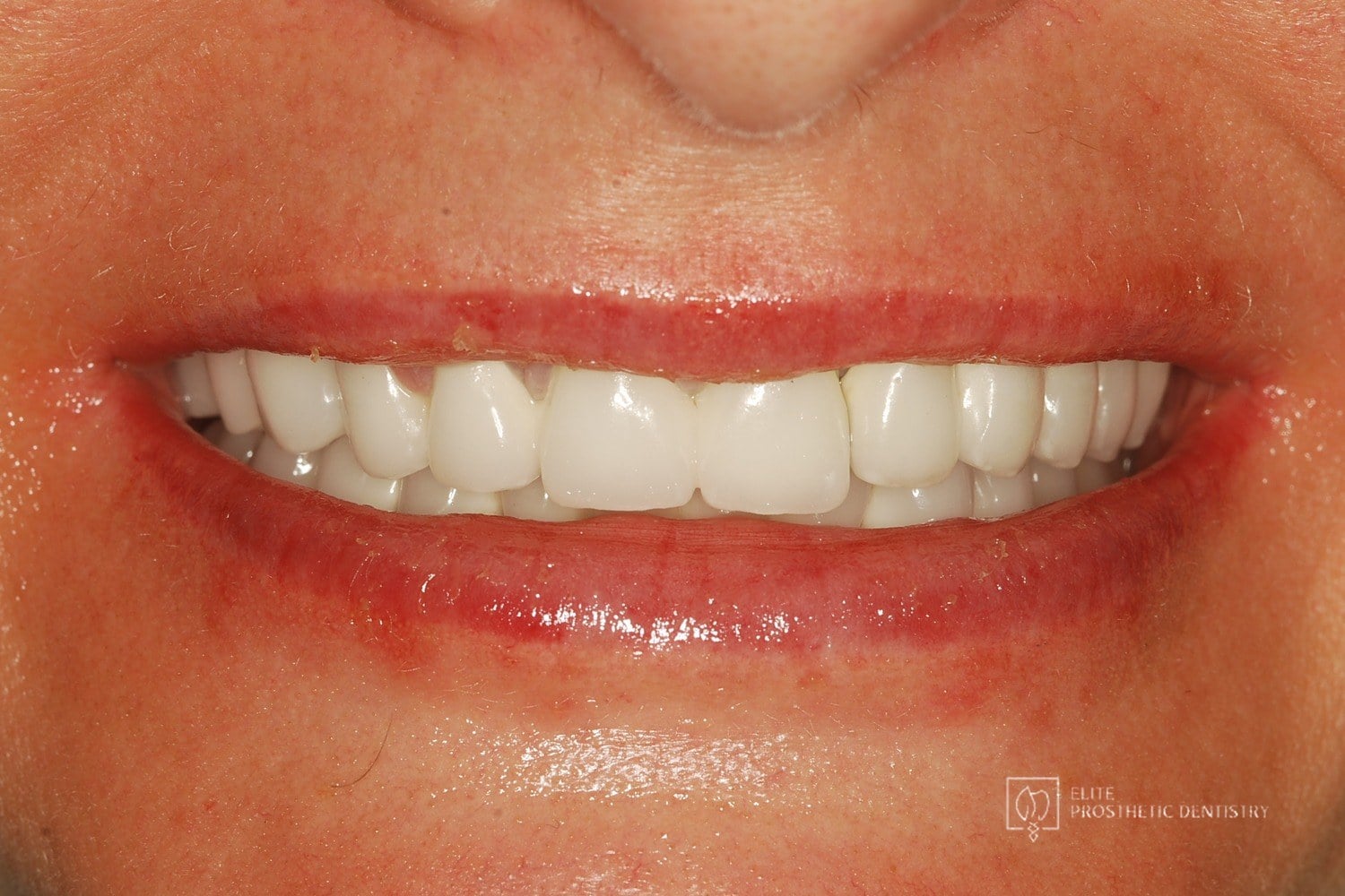
The patient presented with throbbing upper-molar pain, long-standing infections, multiple root canals and unfinished prior treatment, generalized sensitivity, and several hopeless teeth.
 Before
  After
After How Failing Lateral Incisor Implants Were Replaced with Custom Zirconia Bridges After Severe Bone Loss Made Replacement Implants Impossible
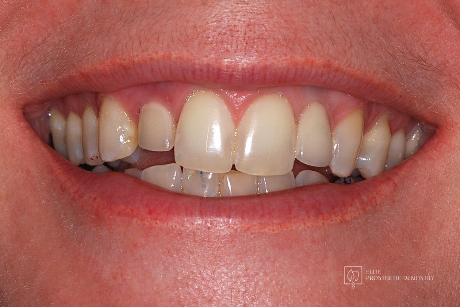
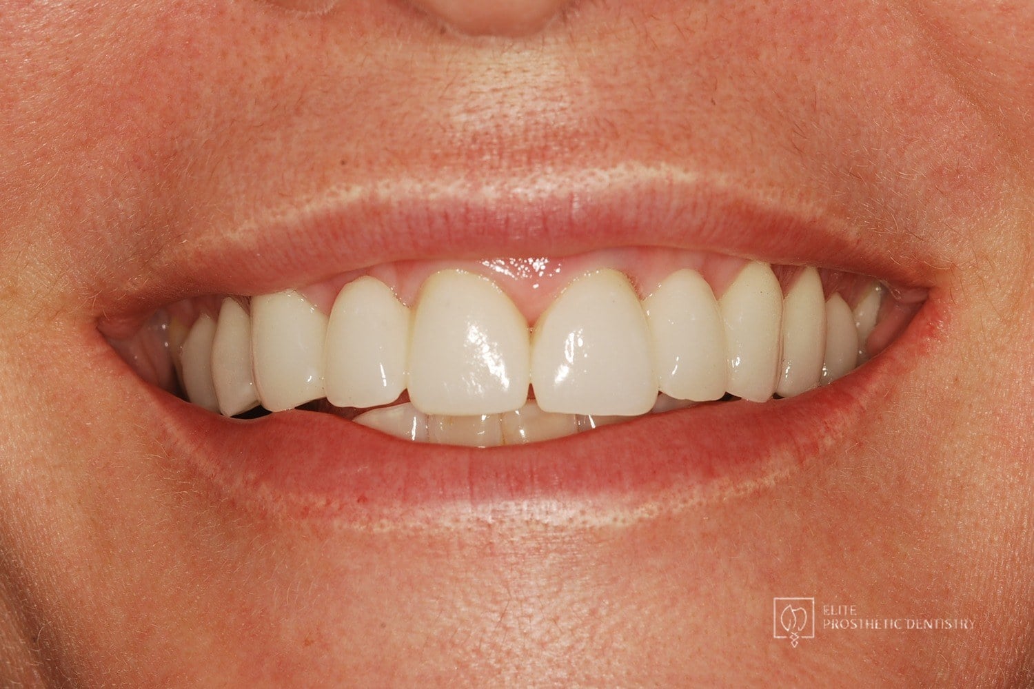
Comprehensive examination including CBCT imaging revealed significant bone loss surrounding implants that were placed outside the buccal plate, most likely due to placement without a CBCT-generated surgical guide. The implants were determined non-salvageable, and the alveolar ridge was too narrow to support replacement implants without extensive reconstruction.
 Before
  After
After How Aging, Opaque Restorations Were Replaced with Customized Ceramic Restorations Designed for Long-Term Natural Esthetics
The existing restorations appeared opaque, worn, and unnatural over time, affecting both confidence and overall smile harmony.